Hemoglobin and Anemia
Hemoglobin is a vital protein in red blood cells responsible for transporting oxygen and carbon dioxide in the body. Learn about its structure, function, synthesis regulation, and factors affecting its activity.

Download Presentation

Please find below an Image/Link to download the presentation.
The content on the website is provided AS IS for your information and personal use only. It may not be sold, licensed, or shared on other websites without obtaining consent from the author.If you encounter any issues during the download, it is possible that the publisher has removed the file from their server.
You are allowed to download the files provided on this website for personal or commercial use, subject to the condition that they are used lawfully. All files are the property of their respective owners.
The content on the website is provided AS IS for your information and personal use only. It may not be sold, licensed, or shared on other websites without obtaining consent from the author.
E N D
Presentation Transcript
Objectives Determination of Glucose-6 phosphate dehydrogenase activity and anima diagnosis Determination of hemoglobin S which diagnose sickle cell anemia Measure the level of hemoglobin
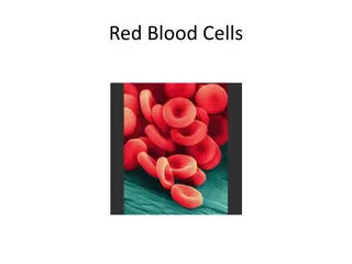
HEMOGLOBIN Hemoglobin is the protein molecule in red blood cells that carries oxygen from the lungs to the body's tissues and returns carbon dioxide from the tissues back to the lungs.
HEMOGLOBIN Hemoglobin (Hb) is a porphyrin iron (II) protien in RBCs that transport oxygen from the lungs to the rest of the body and carbon dioxide back to the lungs. Hb is made up of 4 subunits of globin protein , with a heam (iron containing group).
HEMOGLOBIN The circulation blood of normal adult contain about 750 g of Hb and of this about 7 8 g are degraded daily. This amount has to be newly synthesized each day because: The globin part of Hb can be reutilized only after catabolism into its constituent amino acid. The free heam is broken down into bile pigment which is excreted. Iron alone is reutilized in the synthesis of Hb.
Regulation of Hb Synthesis: Hb synthesis is stimulated by anoxia or hypoxia, whether due to oxygen deficiency or due to anaemia. Anoxia: means a total depletion in the level of oxygen, an extreme form of hypoxia or "low oxygen There is a strong evidence that the marrow response to the stimulus of hypoxia is dependent upon erythropoietin. Erythropoietin is a glycoprotein hormoneformed in kidney in response to decrease oxygen carrying capacity (hypoxia or anoxia), in order to stimulate the erythropoiesis
Regulation of Hb Synthesis: Tissue hypoxia Kidney secrete erythropoietin into blood Return to homeostasis when oxygen is delivered to kidney , this cause negative feedback inhibition to stop the secretion of erythropoietin Increase erythropoiesis Increase number of RBC Increase oxygen carrying capacity
THE ROLE OF SOME FACTOR AFFECTING ON THE NATIVE OF HAEMOGLOBIN: factor affecting on the native of hemoglobin: Glucose -6- phsphatase dehydrogenase (G6PD) Vitamins and cofactor Trace metals Only copper and cobalt are known to play a role . (Copper is playing a role in the absorption of iron while Cobalt is essential constituent of vitamine B12 (Cobalamin) ) Biotin (B7), pantothenic acid (B5), folic acid (B9), coenzyme A and pyrodixal phosphate are essential for haem synthesis .
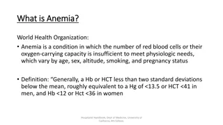
ANEMIA : It is in general decrease in the amount of RBC or the normal amount of Hb in blood.It can also be defined as a lowered ability of the blood to carry oxygen.
Iron-deficiency anemia: Deficiency of iron is essentially due to blood loss with failure to replace the iron stores because of : Dietary deficiency or Increase requirement or Defective absorption. Megaloblastic Anemia: This may be due to deficiency of folic acid or cobaltamin (Vit. B12) RBC membrane defects: In this condition there is a defect of the erythrocyte membrane and an abnormality in the soduim pumps. The best-known disorders are hereditary spherocytosis and hereditary elliptocytosis.
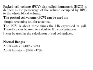
ESTIMATION OF BLOOD HAEMOGLOBIN: Principle: The ferrouse (Iron II) in each haem in RBC is oxidized by ferricyanide to Fe(III)- methaemoglobin . A cynide group (CN-) is then attached to the iron atom (because it is positively charge) by reaction with KCN to give the brown cyanomethamoglobin (stable) which can be estimated quantitatively Normal Hb conc.: for men: 14 - 18 g/dl, for women : 12 - 16 g\dl Level of Hb is associated with polycythemia and dehydration Level of Hb is associated with aneamia
Pipette into clean dry test tubes Test Blank Hemoglobin reagent 2 ml 2 ml Blood sample 0.01 ml ( 10 l) _____ Mix, allow to stand at room temperature for 3 min and read the absorbance at 540 nm against hemoglobin reagent Hb conc (g/dl) = 29.4 x Abs of test
2-QUANTITATIVE DETERMINATION OF G6PD DEFICIENCY IN HEMOLYSED RBC SAMPLE Objectives: Quantitative determination of glucose 6-phosphate dehydrogenase (G6PD) activity in erythrocytes (hemolysate).
GLUCOSE-6 DEHYDROGENASE IMPORTANCE IN RBC RBCs are constantly challenged by oxidants (free radicals) generated by the conversion of oxyhaemoglobin to deoxyhaemoglobin and by peroxides generated by phagocytosing granulocytes. G6PD is an enzyme required to protect cells from oxidation which will cause damage.
GLUCOSE-6 DEHYDROGENASE IMPORTANCE IN RBC It is responsible for the conversion glucose in the pentose phosphate pathway (PPP) to form 6- phosphogluconate , this pathway provide NADPH which is used to produce reduced glutathione (GSH). GSH is necessary for cell integrity by neutralizing free radicals that cause oxidative damage.
GLUCOSE-6 DEHYDROGENASE DEFICIENCY Normal RBCs can increase generation of NADPH in response to oxidative stress; this capacity is impaired in patients with G6PD deficiency. Failure to withstand oxidative stress due to G6PD deficiency,leads to decreased level of NADPH ,therefor Hb is oxidized by free radicals to met-Hb, which aggregates together causing hemolysis. Oxidative stress can result from infection and from chemical exposure to medication e.g. antimalarial drug, and certain foods e.g., fava beans
PRINCIPLE Erythrocytes are lysed (by saponin) and their content is released Glucose + NADP+ G6PD 6-Phosphogluconate + NADPH + H+ The rate of formation of NADPH is a measure of the G6PDH activity and it can be followed by means of the increase in the Absorbance at 340 nm. Note: A red cell hemolysate is used to assay for deficiency of the enzyme, while serum is used for evaluation of enzyme elevations.
METHOD Pipette into clean and dry test tubes Reagent G6PDH Buffer NADP reagent Hemolysate Volume 3 ml 100 l 50 l Mix and incubate for 5 min at 25 C, the add G6PDH Substrate 50 l Mix and read absorbance every min for 3 min against distilled water and calculate A/min
RESULTS A/min=[(A3-A2)+(A2-A1)]/2 Time Abs 340 nm 1 min A1 2 min A2 3 min A3
CALCULATIONS G6PD Activity in mU/erythrocytes/ml of blood ( P )= A/min x 30868 Note: If the erythrocytes count per ml of blood is 5 X 10⁹ Then the G6PD activity in mU/ 109 cells = P/5
3-QUALITATIVE DETERMINATION OF HEMOGLOBIN S (HBS) IN BLOOD. Objectives: Qualitative determination of hemoglobin S (HbS) in blood using a phosphate solubility method.
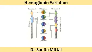
TYPES OF HEMOGLOBINS There are hundreds of Hb variants, and the most common are: Hemoglobin A It is normal hemoglobin that exists after birth and consist of ( 2 2). In normal adult 95% of Hb is present as HbA Hemoglobin A2 It is a minor component of the hemoglobin found in red cells after birth and consists of ( 2 2) less than 3% of the total red cell hemoglobin. Hemoglobin F Hemoglobin F is the predominant hemoglobin during fetal development and consists of ( 2 2).
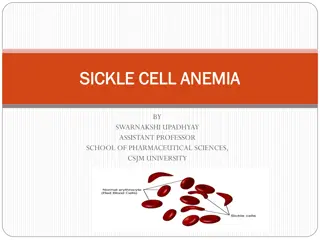
EXAMPLE OF AN ABNORMAL HB Hemoglobin S (HbS) The alpha chain is normal, while the beta chain is mutated, giving the molecule the structure, 2 S2. A point mutation in the Hb gene is responsible for the sickling of RBCs seen in sickle cell anemia .The abnormality is due to Substitution of non polar valine for a charged Glutamic acid in position 6 in the chain .
HbS can be inherited in the homozygous state (S/S) produce sickle cell anemia , or in heterozygous (A/S) ,also called sickle cell trait, usually don t exhibit symptoms of the sickle cell anemia disease (unless under extreme hypoxia). Individuals with HbS will be at high risk when exposed to conditions of low oxygen tension such as surgery, high altitude or athletics which may results in serious and fatal clinical complications.
PRINCIPLE Erythrocytes are lysed (by saponin) and the released hemoglobin is reduced (by dithionite) in phosphate buffer. Reduced HbS is characterized by its very low solubility So that in the presence of HbS, the solution become turbid and the lines behind the test tube will not be visible while, if no HbS was present the clear solution will permit the lines to be seen through the test tubes.
METHOD Pipette into clean dry test tube Reagent Sickling solution Patient sample (whole blood) Mix by inversion and allow stand at room temperature for 5 to 10 min Read the test by holding the test tube approximately 3 cm in front of a lined scale on the card. Volume 2 ml 0.02 ml (20 l)
_ +


 IN")


")