Hemolytic Anemia: Definition, Epidemiology, Etiology, and Clinical Features
Hemolytic anemia is a condition characterized by the destruction of red blood cells, leading to various symptoms such as fatigue, jaundice, and splenomegaly. The etiology ranges from congenital causes like hereditary spherocytosis to acquired causes like autoimmune hemolytic anemia. Diagnosis involves blood tests and imaging studies to identify underlying factors contributing to the disease.

Download Presentation

Please find below an Image/Link to download the presentation.
The content on the website is provided AS IS for your information and personal use only. It may not be sold, licensed, or shared on other websites without obtaining consent from the author.If you encounter any issues during the download, it is possible that the publisher has removed the file from their server.
You are allowed to download the files provided on this website for personal or commercial use, subject to the condition that they are used lawfully. All files are the property of their respective owners.
The content on the website is provided AS IS for your information and personal use only. It may not be sold, licensed, or shared on other websites without obtaining consent from the author.
E N D
Presentation Transcript
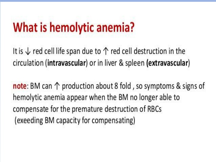
HEMOLYTIC ANAEMIA Definition:- Anemia develops in situations were there is a reduction in RBCs life span due to increase in RBCs destruction associated with failure of compensatory marrow hematopoiesis . Epidemiology :- not common disease , the incidence varying 0.8:1000,000 ---3:1000,000 / year both males & females are equally affected , no definite age of occurrence .
A. Etiology & Causes:- A- CONGENITAL (hereditary) : 1- Membrane abnormality :- hereditary. spherocytosis, hereditary elliptocytosis. 2-Hemoglobinopathies :- Thalassemia, sickle cell disease. 3- Red cell enzymopathies :- glucose -6-PD def. pyruvate kinase def. B- AQUIRED : 1- Immune :- AIHA ( warm Ab , cold Ab ). alloimmune(HDN , RBCs transfusion incompatibility).
2- Non-immune :- a- Mechanical :- artificial cardiac valves, burns, microangiopathic( sepsis, TTP ). b- Infectiuos :- malaria, clostridium welchi. c- Drugs :- penicillins, quinidin. d- Chemicals. e- Dyserythropoietic :- paroxysmal nocturnal hemoglobinuria.
Pathphysiology :- depends on the cause ? eg. AIHA , RBCs will be coated by antibodies which is recognised by RES, and will be hemolysed Clinical Features :- Symptoms :- asymptomatic fatigue , headaches , loss of conc. palpitation , SOB , angina , HF. abdominal pain (GB stones , spleenic infarction). urinary symptoms red or dark brown pallor , jaundice splenomegally hepatomegally LAP. clinically :-
Investigation : CBC PBF reticutocytes & ESR. LFT : including total billirubin D & I , LDH. S. ferritin , RFT & coomb s test (antiglobulin test ) D & I. S. Haptoglobin . Radilogical assessment CXR & ultrasound abdomen, C.T. scan or MRI & Echo. Urine : hemoglobinuria , urobilinogenuria .
Treatment : According to etiological cause - supportive & symptomatic. - Folic acid. - Corticosteroids. - Blood transfusion. - Surgical.
Hereditary Spherocytosis Hemolytic anemia occurs secondary to RBCs membrane defect , inherited as autosomal dominant , presents at any age , both sexes are affected. Clinically : pallor, jaundice, mild to moderate splenomegaly. leg ulceration .
Pathphysiology : The abnormal RBCs lysis is caused by a defect in cytoskeleton ; involves a partial deficiency of Spectrin , the spherical shape has two effects on RBC *trapped in the spleen (shorten life span ) . * the abn.memb. Also increases the permeability of sodium .
Symptoms : - Anemia (compensated to symptomatic). - Gallstones with cholecystitis. The clinical course may be complicated by crisis. - Hemolytic crisis associated with infection. - Megaloblastic crisis following folic acid. deficiency especially in pregnancy. - Aplastic crisis associated with parvovirus infection.
Investigations: - CBC & PBF : spherocytes ,reticulocytosis. - Increased total bilirubin (uncojugated) , high s. LDH ,+ve. Urinary urobilinogen. - (- ve. )Coombs test. Treatment : - Folic acid 5 mg/day life long . - Blood transfusion . - Splenectomy .
Spherocytes Reticulocytes
Autoimmune Hemolytic Anemia : Anemia occurred as a result of presence of RBCs autoantibodies which will coat the RBCs leading to their destruction, it is either warm antibody (IgG) or cold antibody (IgM). Occurs either idiopathic or associated with connective tissue diseases or lymphoma or other lymphoproliferative disorders, Drugs .
Warm autoimmune H. A. IgG binds RBCs in 37 c ,will be hemolysed it accounts 80% of cases, occurs at all ages but common in middle ages and females. clinical features : - symptoms of anemia . - pallor + jaundice . - splenomegaly .
Investigations : - CBC & PBF ( spherocytes , reticulocytosis). - Unconjugated hyperbilirubinemia. - Raised s. LDH levels , reduced haptoglobulins - +ve direct coomb s test. - NB: connective tissue diseases and lymphoma should be excluded.
Treatment : - Prednisolone 1mg /kg + folic acid . - Splenectomy. - Rituximab (anti CD20) . - Immunosuppression ( cyclophosphamide , azathioprine , ciclosporine ) .
Cold Autoimmune H. A. : RBCs are coated by IgM antibodies in 32 c represents 20 % of cases , commoner in elderly patients. either idiopathic or occurs in association of lymphoma , infectious mononucleosis or mycoplasma infection . Clinical presentation : - Pallor, +/- jaundice, splenomegaly. - Acrocyanosis ; painfull, blue fingers, toes, nose, ears on exposure to cold.
Investigation : -CBCs & PBF (agglutination of RBCs) - other inv . Same as in warm AIHA . Treatment : - Folic acid 5mg. - Blood trasfusion. - Prednisolone. - Rituximab. - Treat the underling cause.