Hemorrhagic Fever with Renal Syndrome Overview
Learn about Hemorrhagic Fever with Renal Syndrome caused by hantavirus, its etiology, epidemiology, transmission, and clinical manifestations. Understand the structure of the virus, sources of infection, and prevention strategies in this detailed guide from the Department of Infectious Diseases at Southern Medical University.

Download Presentation

Please find below an Image/Link to download the presentation.
The content on the website is provided AS IS for your information and personal use only. It may not be sold, licensed, or shared on other websites without obtaining consent from the author. If you encounter any issues during the download, it is possible that the publisher has removed the file from their server.
You are allowed to download the files provided on this website for personal or commercial use, subject to the condition that they are used lawfully. All files are the property of their respective owners.
The content on the website is provided AS IS for your information and personal use only. It may not be sold, licensed, or shared on other websites without obtaining consent from the author.
E N D
Presentation Transcript
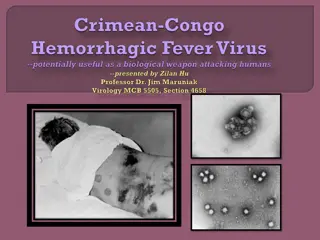
HEMORRHAGIC FEVER WITH RENAL SYNDROME Zhihua Liu Ph.D.&M.D. zhihualiu@126.com Department of Infectious Diseases Nanfang Hospital
OUTLINE Etiology Epidemiology Pathogenesis and Pathology Clinical Manifestations Laboratory Examinations Diagnosis Treatment Prevention Department of Infectious Diseases, Southern Medical University
Introduction Hemorrhagic fever with renal syndrome, also known as epidemic hemorrhagic fever Caused by hantavirus from family Bunyaviridae Carried and transmitted by rodents, especially wild rats Clinically characterized by fever, hemorrhage and renal disorders, usually progresses through five phases, i.e. febrile, hypotensive, oliguric, diuretic and convalescent phases Department of Infectious Diseases, Southern Medical University
Etiology Hantavirus: Classification: Bunyaviridae family Morphology: spherical, 80-120nm in diameter Genome: (-) single strand RNA Tri-segmented genome large: RNA dependent RNA polymerase medium: viral glycoproteins small: nucleocapsid protein Department of Infectious Diseases, Southern Medical University
ETIOLOGY Structure Viral envelop: lipid bilayer+ viral surface protein Nucleocapsid Nucleocapsid protein Viral genome RNA polymerase Department of Infectious Diseases, Southern Medical University
EPIDEMIOLOGY Source of infection Rodents: mice or rats Other vibrate animals: cats, dogs, pigs Human: human-to-human transmission? Department of Infectious Diseases, Southern Medical University
EPIDEMIOLOGY Route of transmission Contact transmission contact with infected animal excreta rodent bite skin breaches Respiratory tract Aerosolized rodent excreta, urine, feces, saliva Alimentary tract food or water contaminated by infected rodents Verticle transmission (spread from mother to fetuses) rare event, not important in epidemiology Department of Infectious Diseases, Southern Medical University
Transmission of Hantaviruses Chronically infected rodent Horizontal transmission of infection between same species by contact Virus is present in aerosolized excreta, particularly urine Secondary aerosols, mucous membrane contact, and skin breaches are also a consideration Department of Infectious Diseases, Southern Medical University
EPIDEMIOLOGY Susceptibility Universal susceptibility Stable and persistant immunity Subclinical infection:2.5%-4.3% Department of Infectious Diseases, Southern Medical University
EPIDEMIC FEATURE Epidemic features Geographic distribution Asian and European countries China is one of most seriously affected countries 40,000~60,000 cases annually Seasonal Distribution: Nov.-Jan., Mar.-Jun. Distribution of population: age:20-40 years old gender: male>female, M:F 2:1-3:1 occupation: farmer, worker in forest, soldier Department of Infectious Diseases, Southern Medical University
Pathogenesis Pathogenesis of disease 1. Direct injury of virus Viremia toxic symptoms Bone marrow cells-- injury Endothelial cells-- injury Department of Infectious Diseases, Southern Medical University
Pathogenesis 2. Immunity injury allegic reaction of type III immune complex allegic reaction of type , T-cell response: Cytotoxic T Lymphocyte (CTL) Cytokine and medium injury IL-1, TNF , IFN Department of Infectious Diseases, Southern Medical University
Pathophysiology of symptoms Shock hantavirus injury of blood vessel vascular permeability exudation of plasma effective blood volume shock Department of Infectious Diseases, Southern Medical University
Pathophysiology of symptoms Hemorrhage Vascular injury Thrombocytopenia diffuse intravascular coagulation(DIC) Platelet dysfunction Department of Infectious Diseases, Southern Medical University
Pathophysiology of symptoms Renal failure Glomerular filtration rate (GFR) Immunity injury of kidney Cast obstruction in renal tubules Interstitial edema pressing renal tubules Department of Infectious Diseases, Southern Medical University
Pathology Basic pathologic lesion Extensive lesion of the systemic small blood vessels Intensive capillary engorgement Diapedesis of erythrocytes Focal hemorrhages Altered capillary permeability Gross pathologic lesions Retroperitoneal edema Diffuse hemorrhage in the right atrium of the heart Severe congestion, hemorrhage and infarct-like necrosis in the renal medulla Hemorrhage and necrosis in the anterior lobe of the pituitary gland Department of Infectious Diseases, Southern Medical University
Clinical manifestations Incubation period: 2-7 weeks, average 7-14 days Five phases: Febrile phase Hypotention-shock phase Oliguric phase Diuretic phase Convalescent phase Department of Infectious Diseases, Southern Medical University
Clinical course and symptoms of HFRS Department of Infectious Diseases, Southern Medical University
Clinical manifestations Febrile phase: fever, suffusion and bleeding, renal impairment Fever:3~7 days, high, unremitting Three pains: headache, lumbago (backache), orbital pain Three reds: conjunctival suffusion; flush over face; flush over neck and upper chest 1. Department of Infectious Diseases, Southern Medical University
Clinical manifestations Hemorrhage: Mucosa: conjunctivae, Palate: petechiae Skin: axillary folds, chest and back petechiae Internal organs Exudative edema: Chemosis; eyelid edema; Renal injury: proteinuria, hematuria or cast Department of Infectious Diseases, Southern Medical University
male, 34 years old, 2 days after illness, hemorrhage in conjunctiva of left eye Department of Infectious Diseases, Southern Medical University Chemosis
female skin of chest and abdomen 31 years old 4 days after illness, petechiae in the Department of Infectious Diseases, Southern Medical University
Membranoid substance in urine Severe kidney injury Department of Infectious Diseases, Southern Medical University
Clinical manifestations 2.Hypotension-shock phase: 4~6 days after illness Mild case: transient fall in blood pressure Moderate and severe case: hypotension and shock persists for 1-3 days Systolic pressure decreases:<90mmHg Cold and moist skin and extremities Pulse pressure narrows Department of Infectious Diseases, Southern Medical University
Clinical manifestations 3. Oliguric Phase: about 5~8 days after illness,last 2~5days Acute kidney injury, Oliguria: urine volume in 24h <500ml Anuria: urine volume in 24h <50ml Uremia Symptoms of digestive tract: anorexia, nausea,vomiting, diarrhea Symptoms of nervous system headache, lethargy,dysphoria BUN increases, electrolyte abnormalities, acidosis Hemorrhage manifestations become more prominent Department of Infectious Diseases, Southern Medical University
Clinical manifestations Disturbance of water and electrolyte balance Hyperkalemia, hyponatremia, exudative edema chemosis, edema of eyelid, ascites High blood volume syndrome Venous engorgement Pulse enlargement Pulse pressure increase Severe edema (heart failure, pulmonary edema) Department of Infectious Diseases, Southern Medical University
Clinical manifestations 4. Diuretic Phase: 9-14 days after illness, lasts 7~14 days Diuresis: urine volume >2000ml/24h Transitional phase urine volume:500~2000ml/24h Early period of diuresis 2000ml~3000ml/24h, azotemia symptom Late period of diuresis >3000ml/24h dehydration hyponatremia, hypokalemia secondary infection Department of Infectious Diseases, Southern Medical University
Clinical manifestations 5. convalescent phase (1-3 months) Urine volume <2000ml/24 hours Department of Infectious Diseases, Southern Medical University
Clinical types Mild type Moderate type Severe type Dangerous severe type Non-typic Department of Infectious Diseases, Southern Medical University
Laboratory findings Blood routine examination Leukocytosis: WBC 15~30 109 heteromorphic lymphocyte >15% Thrombocytopenia Urine routine examination Proteinuria Hemoturia RBC Cast Membranoid substance Department of Infectious Diseases, Southern Medical University
Laboratory findings Serological examination Specific antigen serum, WBC, urine Direct immuofluorescence Specific antibody: IgM antibody, IgG antibody Enzyme immunoassay (EIA) Indirect flurescent assay (IFA) Pathogenic examination Isolation of virus: difficult and time consuming RT-PCR: viral RNA early diagnosis of Hantavirus infection Department of Infectious Diseases, Southern Medical University
Viremia and antibody response in human hantavirus infection Department of Infectious Diseases, Southern Medical University
DIAGNOSIS Epidemiologic data Living or traveling in epidemic region Contact with rodent excreta Clinical features: Three main symptoms Five phases Lab findings Leukocytosis, thrombocytopenia, high percent heteromorphic lymphocyte proteinuria Specific antibody IgM antibody 1:20 positive IgG antibody 1:40 positive Department of Infectious Diseases, Southern Medical University four fold rise
Treatment The principle of therapy Early diagnosis Recognizing timely Treatment rapidly and in site Hospitalized and kept in bed Avoid movement by vehicles Monitored intensively Antipyretic drug Supportive therapy Department of Infectious Diseases, Southern Medical University
Treatment Febrile phase Controlling infection: ribavirin, as early as possible Relieve permeability of blood vessel Hemostatic Vitamin C Mannitol Alleviate toxic symptom: High fever: physical cooling Toxic symptom: Glucocoticoid: dexamethasone,hydrocortisone Department of Infectious Diseases, Southern Medical University
Treatment Hypotensive phase Supplement of blood volume Early, fast, suitable volume Crystalloid solution plus colloidal solution crystalliod solution: normal saline solution, compound sodium acetate colloidal solution: plasma, human albumin crystalliod: colloidal 3~4:1 Correction of acidosis 5% sodium bicarbonate (NaHCO3) Vaso-active agent Dopamine: 100~200mg/L Department of Infectious Diseases, Southern Medical University
Treatment Oliguric peroid Principle of treatment: Stabilization of internal environment Promoting diuresis hemodialysis Treating and preventing complications Department of Infectious Diseases, Southern Medical University
Treatment Stabilization of internal environment keeping the water, electrolytes and acid-base in the body in the physiology range Input solution should be limited at a rational extent output volume plus 500-800ml Department of Infectious Diseases, Southern Medical University
Treatment Diuresis started 12~24 hours after the blood pressure become stable Loop diuretics: furosemide, bumetanide Dialysis hemodialysis- the most effective therapeutic methods for acute renal failure indications for hemodialysis: Oliguria > 5 days or anuria> 2 days Hypervolemia syndrome with pulmonary edema, cerebral edema Hyperkalimia Disease progress rapidly Department of Infectious Diseases, Southern Medical University
Treatment Diuretic phase electrolytes unbalance Dehydration Infection Convalescent phase Nutrition, functional exercise Department of Infectious Diseases, Southern Medical University
Prevention Rodent control in highly endemic areas Avoid contacts with the host rodents Vaccine Inactived hantavirus vaccines used in China Department of Infectious Diseases, Southern Medical University
Summary HFRS is a group of diseases with fever, hemorrhage and renal injury Pathogen: Hantaviruses HFRS consists of five phases: Febrile, hypotention, oliguric, diuretic, convalescence Department of Infectious Diseases, Southern Medical University
THANK YOU! Department of Infectious Diseases, Southern Medical University