Hog Cholera - A Veterinary Perspective
Hog cholera, also known as swine fever, is a highly contagious viral disease affecting pigs. Learn about its epidemiology, control measures, etiology, and more. Discover ways to prevent and manage outbreaks to safeguard pig populations.

Download Presentation

Please find below an Image/Link to download the presentation.
The content on the website is provided AS IS for your information and personal use only. It may not be sold, licensed, or shared on other websites without obtaining consent from the author.If you encounter any issues during the download, it is possible that the publisher has removed the file from their server.
You are allowed to download the files provided on this website for personal or commercial use, subject to the condition that they are used lawfully. All files are the property of their respective owners.
The content on the website is provided AS IS for your information and personal use only. It may not be sold, licensed, or shared on other websites without obtaining consent from the author.
E N D
Presentation Transcript
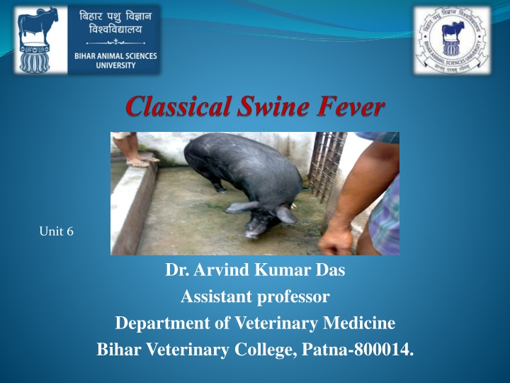
Unit 6 Dr. Arvind Kumar Das Assistant professor Department of Veterinary Medicine Bihar Veterinary College, Patna-800014.
Introduction Also called Hog Cholera / Swine fever Highly contagious and febrile viral disease of swine Disease may occur from per-acute and acute to sub-acute and chronic, and can last for several weeks or even months. Notifiable to OIE Naturally susceptible: Only the domestic pig and wild boar No infection to human Morbidity and mortality : Zero to 100 % Mortality high in young ones
Epidemiology World wide Endemic in parts of Latin America, Caribbean islands & pig producing countries of Asia First notice as an animal disease in early 19th century in USA Epidemic: In Minnesota in 1913 South Africa in 1918 Netherland in1997-98: 429 herds infected, 700,000 pigs culled
Policies adopted to Control Controlled by: Early detection and control to spread High Risk Period: Shortest time between introduction of virus and detection of outbreak with clinical symptoms Restricted movements Culling of infected and suspected pigs Total 12 million pigs slaughtered Australia, New Zealand, Canada and USA are free from CSF Many country banned the pig meat feeding
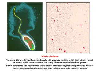
Etiology Small enveloped RNA virus Genus: Pestivirus Family: Flaviviridae Antibody discrimination tests must to differentiate Only one serotypes with minor antigenic variability, divided into three major genotypes, including numerous sub-genotypes
Close Relationship Antigenically related to BVDV of cattle and Border disease virus of sheep BVD CSF Border disease
Characters of virus Moderately fragile No persistent in environment- No spread from long route by airborne infection Survive prolonged in moist protein rich medium, meat, tissues, body fluids if frozen or chilled Survive several years in frozen pig meat and months in chilled meat Inactivated by cooking
Source of Infection Main source: wild boar population or domestic Pigs (live or uncooked pig product) Wild boar populations may harbour virus; domestic pigs in the affected area are at a high risk Spread by: Movement of infected pigs Contaminated garbage Accidental introduction of virus through illegally imported pig meat.
Transmission Routes : Oral and oronasal via direct or indirect contact Direct: Secretions, excretions, semen, blood Indirect : Contact through premises, implements, vehicles, clothes, instruments, needles and insects Neighbourhood effect during outbreaks in areas of high pig farm density: airborne transmission over short distances Spread by mechanical transmission: Vehicles, equipments and personnel travelling between pig farm and infected area
Tran-placental Transmission The virus can pass from the sows to their offspring Tran-placental infection: Offspring's of pregnant ewes infected with low or moderately virulence strain and recovered at high risk and may be carriers. Not showing symptoms but permanently shed the virus. Important to investigate herds having high level of unexplained reproductive failure and congenital tremor or other abnormalities
Clinical Findings Acute & Chronic Forms Virulence: Severe - High mortality Mild or subclinical Low Poor reproductive performance, congenital neurologic defect in newborn piglet
Severe Acute Form: Clinical Signs Fever (>41 C) until terminal stage when temperature subnormal Inappetence Depression Incubation period 3 to 5 days Death 10 to 20 days after infection Anorexia Constipation followed by diarrhoea
Severe Acute Form.Continued Multifocal hyperaemia and/or haemorrhagic lesions of the skin Conjunctivitis Enlarged, swollen lymph nodes Cyanosis of the skin especially of extremities (ears, limbs, tail, snout) Transient constipation followed by diarrhoea Vomiting (occasional)
Severe Acute Form.Continued Lethargy Dyspnoea, coughing Ataxia, paresis and convulsion Pigs huddle together Mortality in young pigs can approach 100%
Chronic form Dullness, capricious appetite, pyrexia, diarrhoea for up to 1 month Ruffled appearance of pigs Growth retardation Poor reproductive performance may be the only sign in some breeding herds infected with less virulent strains Apparent recovery with eventual relapse with anorexia, depression, fever, and progressive loss of condition and death within 3 months
Lesions Enlarged hemorrhagic lymph nodes are common Generalized vasculitis Vasculitis in CNS lead to incordination or convulsions Hemorrhages and cyanosis in skin especially in extremities Generalized erythema Severe tonsillitis with necrotic foci sometimes occurs
Lesions Necropsy: Widespread petechial or echymotic hemorrhages in skin, lymph nodes, epiglottis larynx kidneys, spleen, bladder and rectum, specially in spleen Multifocal infarction of the margin of the spleen is characteristic: nearly pathognomonic but occurs infrequently with currently circulating strains Nonsuppurative encephalitis with vascular cuffing Histologically: Atrophy of thymus, lymphoid depletion Button shaped ulcers in intestine particularly near ileocecal junction.
Tests Samples Tissues of tonsil Maxillary or submandibular lymph node Mesenteric lymph node Spleen Ilium and Kidney Whole blood with EDTA for virus isolation Clotted blood for serologic test
Diagnosis Virus isolation o Isolation in PK-15 cell line culture o Demonstration by an immunostaining method (FAT or immunoperoxidase) RT-PCR Detects infected animals early during the incubation period and for a longer period of reverse transcriptase polymerase chain reaction (RT-PCR) techniques time in cases where the pigs recover. In this test virus nucleic acid detection is done. Fluorescent antibody test (FAT) It is a rapid test that can be used to detect CSFV antigen ELISA: Also detect antigen but low sensitivity only screened herd level Serological tests Due to the immunosuppressive effect of CSFV, antibodies cannot be detected with certainty until at least 21 days post-infection.
Differential Diagnosis With other febrile hemorrhagic diseases of pigs African swine fever Bacterial septicemias (eg. Salmonellosis, Erysipelas etc) Anticoagulant poisoning (coumarin type) Hemolytic disease of newborn Low reproductive performance and congenital tremors differentiated with pseudorabies, parvovirus and BVDV
Prevention and Treatment No specific treatment available. Infected or in contact animal slaughtered and carcasses buried or incinerated Modified live vaccines (MLVs) based on several attenuated virus strains are most widely used, and many of them have proven to be both safe and efficacious. New generations of marker vaccines are also being developed, including a new chimeric Pestivirus vaccine that has been licensed by the European Medicines Agency (EMA). DIVA??