Hydatid Disease: Causes, Symptoms, and Treatment
Hydatid disease, caused by Echinococcus granulosus, is a parasitic infection affecting human tissues, primarily the liver and lungs. Learn about the morphology, geographical distribution, and cyst characteristics associated with this disease. Understand the complexities of hydatid cysts, including their size, shape, and contents. Explore the diagnostic techniques and treatment options available to manage this condition.

Download Presentation

Please find below an Image/Link to download the presentation.
The content on the website is provided AS IS for your information and personal use only. It may not be sold, licensed, or shared on other websites without obtaining consent from the author.If you encounter any issues during the download, it is possible that the publisher has removed the file from their server.
You are allowed to download the files provided on this website for personal or commercial use, subject to the condition that they are used lawfully. All files are the property of their respective owners.
The content on the website is provided AS IS for your information and personal use only. It may not be sold, licensed, or shared on other websites without obtaining consent from the author.
E N D
Presentation Transcript
1. Hydatid disease Echinococcus granulosus 2. Amoebiasis Entamobae histolytica
Definition: it is the presence of hydatid cyst, larval stage of Echinococcus granulosus in the human tissues. The liver is the commonest organ affected (70%) followed by the lungs (20%), then the brain and other organs (10%).
Geographical distribution: cosmopolitan, more in cattle raising countries.
Morphology: 1. Adult: - Size: about 5 mm. - Scolex: globular with 4 suckers and double crown of hooks (similar to T. solium). -Strobila: composed of 3 segments, one immature, one mature and one gravid. - Mature segment: longer than broad. Reproductive organs as Taenia. -Gravid segment: is 1/2 length of the worm, longer than broad. The uterus develops lateral pouches which are full of eggs.
2. Egg: similar to T. solium - Size: 30-40 - Shape: spheroid. - Shell: thick radially striated embryophore - Colour: yellowish-brown. - Contents: hexacanth embryo (onchosphere) 3. Hydatid cyst:a complex cyst composed of daughter cyst inside and may be outside the mother cyst and contains several scolices.
Unilocular hydatid cyst: it is the larval stage of E. granulosus. - Size: 1-10 cm. - Shape: spherical enclosed in a fibrous capsule produced by the host. - The wall of the cyst has 2 layers: (l) Outer laminated non-cellular layer. (2) Inner cellular germinal layer which secretes the laminated layer and produces scolices, brood capsules and daughter cysts.
- Contents: (a) Individual scolices (microscopic). (b) Brood capsules: invagination of the germinal layer from which scolices develop. (c) Daughter cysts: cysts formed of the 2 layers of the mother cyst, giving rise to scolices, brood capsules and even grand daughter cysts. (d) Hydatid fluid. (e) Hydatid sand: detached scolices, brood capsules and daughter cysts that fall in the hydatid fluid are called hydatid sand. (f) Exogenous daughter cysts: a daughter cyst is produced outside the mother cyst by herniation through the fibrous capsule and may separate from it.
Life cycle: - Habitat: small intestine - DH: dogs or other carnivorous animals - IH: herbivorous animals (cattle) and occasionally man - Infective stage to dog: hydatid cyst - Infective stage to man: Eggs - Stages of life cycle: egg, onchosphere, hydatid cyst in (I. H.), adult in (D. H.). -Gravid segments and mature eggs pass in the faeces of definitive host. -When the egg is ingested by the intermediate host, the liberated onchosphere penetrates the intestinal wall into the blood stream to various parts of the body where it develops into a hydatid cyst, causing hydatid disease.
-Infective stage: Echinococcus egg -Diagnostic stage: hydatid cyst, hydatid sand
Mode of human infection: ingestion of eggs of Echinococcusspecies by the following ways: 1-Ingestion of water or vegetable polluted by infected dog's faeces. 2-Handling infected dogs where hair is usually contaminated with eggs. 3-Man is infected with the alveolar cyst during the skinning of foxes to make furs or from collecting strawberries polluted with fox's faeces.
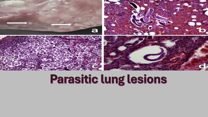
Pathogenicity and clinical picture: depend on the size of the cyst and the organ affected. 1- Hepatic hydatid cyst: indigestion, jaundice and discomfort in the right hypochondrium. 2- Pulmonary hydatid cyst: dyspnea, cough, chest pain and haemoptysis. 3- Cerebral hydatid cyst: symptoms of increased intracranial tension and epilepsy. 4- Osseous hydatid cyst: erosion and spontaneous pathological fracture of long bones. 5- Rupture of the cyst results in anaphylactic shock and transplantation of the germinal layer in other tissues producing secondary cysts.
Geographical distribution: Worldwide distribution especially in tropical areas and poor communities.
Morphology: 1. Trophozoite (Vegetative or growing stage): -Size: 10-60 (average 20 ). -Shape: Irregular outline with finger like pseudopodia and active movement. - Cytoplasm: It is formed of outer clear hyaline, refractile ectoplasm and inner granular endoplasm containing nucleus, food vacuoles, erythrocytes (RBCs), occasionally bacteria, and tissue debris. - Nucleus: It has centrally located fine karyosome and peripheral chromatin dots arranged regularly at the inner side of the nuclear membrane.
2. Cyst: - Size: 10-15 in diameter. - Shape: It is rounded, - Wall: Has smooth refractile cyst wall. - Content: The early cyst contains glycogen vacuoles and 1-4 chromatoid bodies which are sausage-shaped with rounded ends. They are formed of RNA & DNA, and represent stored proteins which are consumed with repeated nuclear division. - Immature cysts may be mono- or bi-nucleated. - Mature cysts contain 4 nuclei formed by mitotic division. - Nuclei are similar to that of the vegetative form.
Life cycle: -Habitat: a. Trophozoite: Inhabits the wall and lumen of the large intestine, with extra-intestinal metastases (liver, lung and brain, etc.). b. Cyst: Inhabits the lumen of the large intestine. - DH: Man - IH: No - RH: Dogs, rats and monkeys. - Infective stage: Mature quadrinucleated cyst - Diagnostic stage: Trophozoite - Stages of life cycle: Trophozoite and cyst
Mode of human infection: 1. Ingestion of mature quadrinucleated E. histolytica cysts in contaminated food or drink, or through infected food handlers. 2. Mechanical transmission by flies and cockroaches. 3. Autoinfection: feco-oral route (hand to mouth contact). - Trophozoite may invade the wall of large intestine by their lytic secretion to invade the host tissues through blood vessels (extra-intestinal invasion).
Pathogenicity: - Entamoeba trophozoites attach themselves to the surface epithelium aided by an enzyme called E. histolytica lectin and start crawling over the mucosa. -Trophozoites secrete cytolytic enzymes; haemolysins and pore-forming enzymes (amoeba pore), which lead to necrosis of epithelial cells with pore formation. - Tophozoites enter to the submucosa through the hole formed in the epithelial layer and continue the process of cytolysis downwards and laterally.
- The lesion is flask shaped or crater like ulcer containing cytolysed cells, mucus and amoeba trophozoites, while amoeba cysts never found in tissues. - Ulcers are more common in the ileocaecal region followed by the sigmoid- rectal region. - Ulcer expansion can penetrate the intestinal wall intestinal perforation with hemorrhage and peritonitis. - Invasion of blood vessels may lead to spread of amoebae causing extra intestinal amoebiasis.