Hypoxia and Cyanosis
Hypoxia is the inadequate supply of oxygen to body tissues, classified into types such as hypoxic hypoxia, anemic hypoxia, stagnant hypoxia, and histotoxic hypoxia. Explore the causes of hypoxia, including inadequate oxygenation of blood in the lungs, pulmonary diseases, and venous-to-arterial shunts. Learn about hypoxic hypoxia, also known as arterial hypoxia, which results from a lack of oxygenation of blood in the lungs.

Download Presentation

Please find below an Image/Link to download the presentation.
The content on the website is provided AS IS for your information and personal use only. It may not be sold, licensed, or shared on other websites without obtaining consent from the author.If you encounter any issues during the download, it is possible that the publisher has removed the file from their server.
You are allowed to download the files provided on this website for personal or commercial use, subject to the condition that they are used lawfully. All files are the property of their respective owners.
The content on the website is provided AS IS for your information and personal use only. It may not be sold, licensed, or shared on other websites without obtaining consent from the author.
E N D
Presentation Transcript
Hypoxia & Cyanosis Color index: Red: important Green: doctor s notes Grey: extra information Pink: found only in female s slides Blue: found only in male s slides Yellow: numbers contact us at: physiologyteam437@gmail.com @physio437 Physiology 437 team work Editing file
objectives: By the end of the lecture you will be able to: By the end of this lecture you should be able to: Define hypoxia and list its various physiological and pathological causes. Define hypo and hyper-ventilation in terms of arterial PCO2 and PO2. Define cyanosis and its clinical presentation Define ventilation/perfusion (V /Q) ratio and its normal values.
Hypoxia vs hypoxemia: Hypoxia is the deficiency of O2 in tissue Hypoxemia is the deficiency of O2in blood Hypoxia Hypoxia is defined as an inadequate supply of oxygen to the body tissues. (deficiency of oxygen in the tissue cells) It can be classified in the following types: 1. Hypoxic hypoxia (arterial hypoxia) e.g. at high altitudes, low O2in air 2. Anemic hypoxia decreased percentage of hemoglobin in blood with respect to age & gender, could be caused by severe loss of blood 3. Stagnant hypoxia (Hypokinetic or Ischemic hypoxia) lack of blood supply 4. Histotoxic hypoxia tissues are unable to utilize O2
Only in males slides. CLASSIFICATION BASED ON THE CAUSES OF HYPOXIA 1. Inadequate oxygenation of the blood in the lungs because of extrinsic reasons a. Deficiency of oxygen in the atmosphere b. Hypoventilation (neuromuscular disorders) 2. Pulmonary disease a. Hypoventilation caused by increased airway resistance or decreased pulmonary compliance b. Abnormal alveolar ventilation-perfusion ratio (including either increased physiologic dead space or increased physiologic shunt) from alveolar damage c. Diminished respiratory membrane diffusion due to increased thickness of membrane e.g. emphysema 3. Venous-to-arterial shunts ( right-to-left cardiac shunts). Damage of septum between atria or ventricles deoxygenated blood in systemic circulation hypoxia
Only in males slides. Cont. 4. Inadequate oxygen transport to the tissues by the blood a. Anemia or abnormal hemoglobin b. General circulatory deficiency c. Localized circulatory deficiency (peripheral, cerebral, coronary vessels) d. Tissue edema e.g. pleural edema impairs diffusion of gases hypoxia 5. Inadequate tissue capability of using oxygen tissue receives O2 but can t use it a. Poisoning of cellular oxidation enzymes this is how cyanide works b. Diminished cellular metabolic capacity for using oxygen, because of toxicity, vitamin deficiency, or other factors.
Hypoxic Hypoxia Hypoxic hypoxia is also known as arterial hypoxia. This is seen when there is a lack of oxygenation of blood in the lungs, which leads to a low PO2 in arterial blood. Since less amounts of Hb is converted to oxy-Hb. The tissues are supplied with blood deficient ( ) in oxygen. Hypoxic hypoxia can occur in the following conditions: High altitude (leading cause) Fluid in the lungs (pulmonary edema) Obstruction in the respiratory passages e.g. obstruction of bronchi Emphysema
Hypoxic Hypoxia Other causes of hypoxic hypoxia: Alveolar hypoventilation Diffusion abnormalities. E.g pulmonary edema & pneumonia Right to left shunt Ventilation-perfusion imbalance (including increased physiological dead space and physiological shunt similar to Venous-to-arterial shunt. Only treatment is through surgery of septum). Alveolar hypoventilation: e.g. Reduced PO2 in inspired air (high altitude). Increased airway resistance. Reduced lung compliance. Compliance is the ease at which the lung can expand Paralysis of respiratory muscles Depressed respiratory centre. Only in male s slides.
Only in males slides. Hypoxic Hypoxia Diffusion abnormalities: Impaired diffusion from alveolar to pulmonary capillary blood can lead to arterial hypoxia. It is seen in conditions like alveolar-capillary block. Ventilation-perfusion imbalance (including increased physiological dead space and physiological shunt): If ventilation and blood flow are mismatched in various parts of the lung, impairment of both oxygen and carbon dioxide diffusion results. Ventilation perfusion imbalance may be caused by uneven ventilation, (e.g. obstructive lung conditions) , or uneven perfusion, (e.g. consolidation of the lung). Right to left shunt: Blood passes from the systemic venous without going through the gas exchanging part of the lungs. This type of hypoxia can be differentiated clinically from other types by giving the subject 100% oxygen to breathe. Hypoxia because of the shunt will not be abolished while in other types PO2 in the arterial system will improve considerably. It s congenital disorder.
Anemic Hypoxia This condition is characterized by decreased oxygen carrying capacity of the blood due to decreased hemoglobin level or abnormal type of hemoglobin which is unable to carry oxygen. Causes: Anemia. Abnormal Hb (Altered haemoglobin formation) e.g. methaemoglobin, sulfhemoglobin, and carboxyhemoglobin. High concentrations of abnormal Hb impair the blood s capacity to carry oxygen Hemorrhagic anemia [Decreased RBC / quality].Quantity could be decreased from trauma or any other loss of blood. While quality could be caused by improper formation of RBCs.* The failure of hemoglobin to carry its normal concentration of oxygen, as in carbon monoxide (CO) poisoning. *Presence of the Nucleus in RBCs is a clear indicator of improper formation
Stagnant Hypoxia Stagnant hypoxia (Hypokinetic or Ischemic hypoxia) occurs in conditions in which there is a decreased rate of blood flow throughout the body or a part of the body (tissue). so more and more oxygen is extracted from the blood, and due to slow circulation less oxygen is carried by the blood at the lung , leading to hypoxia. Causes: It may be caused by congestive heart failure, circulatory shock and arteriosclerosis*. General slowing of circulation (heart failure and shock). Local slowing: vasoconstriction, cold and arterial wall spasm. Arteriosclerosis: decrease of the diameter of a blood vessel due to fat deposition stagnant hypoxia in the hand.
Histotoxic Hypoxia In histotoxic hypoxia the tissues are unable to use oxygen even though plenty of oxygen is available. Caused by: inhibition of the tissue respiration electron transport chain (inhibition of the oxidative enzyme activity) The best example: cyanide poisoning, where tissue cytochrome oxidases are knocked out and tissue is unable to utilize oxygen.
Hypoxia & Clinical features Signs and symptoms of hypoxia : clinical features depend on how fast and how severely partial pressure of O2 is decreased. Chronic hypoxia Fulminant hypoxia Acute hypoxia This occurs very rapidly, within seconds. Unconsciousness occurs in 15-20 seconds and brain tissue death occurs in 4-5 minutes. In acute hypoxia body reflexes are slowed, there may be slurred speech, unconsciousness, coma and death may occur. The symptoms of chronic hypoxia, fatigue, difficulty in breathing, and shortness of breath can occur. Signs of hypoxia : Cyanosis, Tachycardia and Tachypnea (breathing rapidly).
Effects of hypoxia on the body Hypoxia, if severe enough, can cause death of cells throughout the body, but in less severe degrees it causes principally depressed mental activity, sometimes culminating in coma, reduced work capacity of the muscles. At 12,000 feet, drowsiness, lassitude, mental and muscle fatigue, sometimes headache, occasionally nausea, and euphoria. These effects progress to a stage of twitchings or seizures above 18,000 feet and above 23,000 feet in the unacclimatized person, in coma, followed shortly thereafter by death. One of the most important effects of hypoxia is decreased mental proficiency, which decreased judgment, memory,Inability to perform complex calculations and performance of discrete motor movements. If an unacclimatized aviator stays at 15,000 feet for 1 hour, mental proficiency ordinarily falls to about 50% of normal, and after 18 hours at this level it falls to about 20% of normal.
ONLY IN FEMALES SLIDES Treatment of hypoxia Hypercapnia Administration root: Is giving by oxygen therapy in a tent or high oxygen tension mask. Excess of CO2 in body fluid, it usually occurs with hypoxia, PCO2 increases above 52 mmHg, it decreases the PH. Features of hypercapnia : 1. Peripheral vasodilation 2. Sweating 3. Warm extremities and bounding pulse 4. Muscle twitching 5. headache, drowsiness and coma 6. Papilledema ( swelling of optic disc) This is useful in hypoxic hypoxia, but of less value in other types of hypoxia. Histotoxic hypoxia will not benefit from O2 therapy
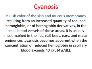
Cyanosis The term cyanosis means blueness of the skin and mucous membrane, and is caused by excessive amounts of deoxygenated hemoglobin in the skin blood vessels, especially in the capillaries. This deoxygenated hemoglobin has an intense dark blue-purple color that is transmitted through the skin. Cyanosis appears whenever the arterial blood contains more than 5 grams of deoxygenated hemoglobin in each 100 millimeters of blood.
CYANOSIS A person with anemia almost never becomes cyanotic because there is not enough hemoglobin for 5 grams to be deoxygenated in 100 milliliters of arterial blood. (Anemia > Less blood cells > Less hemoglobin to be deoxygenated > Cyanosis cannot be achieved due to the lack of hemoglobin and red blood cells). Conversely, in a person with excess red blood cells, as occurs in polycythemia vera, the great excess of available hemoglobin that can become deoxygenated leads frequently to cyanosis, even under otherwise normal conditions. (Polycythemia > More blood cells > More hemoglobin to be deoxygenated > Cyanosis can be achieved due to the excess hemoglobin and red blood cells.)
ONLY IN MALES SLIDES Causesof Cyanosis Inadequate oxygenation of blood in the lungs Presence of an aerated shunt between vessels Other Causes High altitude Obstruction passages Pneumoconiosis (a disease of the lungs due to inhalation of dust, characterized inflammation, coughing, and fibrosis) Emphysema CO-poisoning of respiratory Coarctation of aorta Fallot's tetralogy Moderate cold Diminished blood flow to tissues by
ONLY IN MALES SLIDES Types of cyanosis Local cyanosis: This is seen during decreased blood flow through a part of the body as in Raynaud s disease. In circulation through the upper limb is impaired, it causes local cyanosis. Generalized cyanosis: Generalized impairment of circulation as in the case of heart failure generalized cyanosis. It also occurs in hypoxic hypoxia. leads to this disease,
ONLY IN MALES SLIDES CYANOSIS Dr. Sultan says it s not very important Causes of central cyanosis: Cyanotic congenital heart-disease Fallot tetralogy Tricuspid atresia Ebstein s anomaly Pulmonary arteriovenous fistula Pulmonary diseases Acute pulmonary embolism Pneumonia Atelectasis Chronic Obstructive airway disease Restrictive lung disease Hemoglobin abnormalities Causes of peripheral cyanosis Reduced cardiac output, as in congestive heart failure, mitral stenosis Exposure to cold Arterial obstruction Venous obstruction Raynauds disease Polycythemia vera
ONLY IN FEMALES SLIDES Ventilation perfusion ratio (V/Q) 3;32 It is the ratio of alveolar ventilation to pulmonary blood flow per minute. The alveolar ventilation at rest (4.2 L/min) The pulmonary blood flow is equal to right ventricular output per minute (5L/min) SO, V/Q ratio = 4.2\5 = 0.84 Average V/Q ratio across the lung is 0.8 Ventilation At the apex V/Q ratio = 3 At the base V/Q ratio=0.6 perfusion So the apex is more ventilated than perfused and the base is more perfused than ventilated. During exercise the V/Q ratio becomes more homogenous among different parts of the lung.
ONLY IN FEMALES SLIDES Ventilation/perfusion abnormalities Perfusion Ventilation Ventilation Perfusion Ventilation Perfusion Ventilation perfusion Perfusion Ventilation Q=0
ONLY IN FEMALES SLIDES Ventilation perfusion ratio (V/Q) 3:34 The main function of this ratio is to determine the state of oxygenation in the body. Any mismatch in the ratio can result in hypoxia. When the V/Q ratio is less than normal this is called physiologic shunt (a certain fraction of the venous blood is passing through the pulmonary capillaries without being oxygenated i.e shunted blood). When V/Q is more than normal this is called Physiologic dead space (when the ventilation of some of the alveoli is great but the alveolar blood flow is low, ventilation of these alveoli is wasted).
ONLY IN FEMALES SLIDES Ventilation-Perfusion Lung Scan Lungs Ventilation Perfusion
Abnormalities of the V/Q ratio ONLY IN FEMALES SLIDES In the Upper and Lower regions of the normal lung - Apex V/Q ratio = 3 (moderate degree of physiologic dead space) - Base V/Q ratio= 0.6 ( represent a physiologic shunt). In Chronic Obstructive Lung disease COPD because of bronchial obstruction in some areas and destruction of the alveolar septa in other areas with patent alveoli those people has some areas of the lung exhibit serious physiologic shunt and in other areas serious physiologic dead space. COPD is the most prevalent cause of pulmonary disability today, lung effectiveness as a gas exchange organ may decrease to 10%
SUMMARY Hypoxic Hypoxia : This is seen when there is a lack of oxygenation of blood in the lungs, which leads to a low PO2 in arterial blood. Hypoxia is defined as an inadequate supply of oxygen to the body tissues. Hypoxia types : Anemic Hypoxia : This condition is characterized by decreased oxygen carrying capacity of the blood due to decreased hemoglobin level or abnormal type of hemoglobin which is unable to carry oxygen. Classification Based On The Causes Of Hypoxia : 1-Inadequate oxygenation of the blood. 2-Pulmonary disease. 3-Venous-to-arterial shunts. 4-Inadequate oxygen transport to the tissues. 5-Inadequate tissue capability of using oxygen. 1-Hypoxic hypoxia 2-Anemic hypoxia 3-Stagnant hypoxia 4-Histotoxic hypoxia
SUMMARY Histotoxic Hypoxia : In histotoxic hypoxia the tissues are unable to use oxygen even though plenty of oxygen is available. Stagnant Hypoxia : occurs in conditions in which there is a decreased rate of blood flow throughout the body or a part of the body (tissue). Cyanosis Clinical features : 1-Fulminant hypoxia 2-Acute hypoxia 3-Chronic hypoxia The term cyanosis means blueness of the skin and cause is excessive amounts of deoxygenated hemoglobin in the skin blood vessels, especially in the capillaries. Causes of Cyanosis 1-inadequate oxygenation of blood in the lungs 2-Presence of an aerated shunt between vessels 3-Other Causes Signs of hypoxia : 1-Cyanosis 2-Tachycardia 3-Tachypnea Types of cyanosis 1-Local cyanosis 2-Generalized cyanosis
Females team: Leader: Alanoud Salman Alotaiby Members: Majd AlBarrak Dimah Alaraifi Maha Barakah Maha Alnahdi Sarah AlFlaij Male s team: Leader: Abdulhakim AlOnaiq Members: Rayyan Almousa Abduljabbar Alyamani