Idiopathic Talipes Equinovarus (Clubfoot) and its Characteristics
Explore the clinical features, epidemiological aspects, and diagnostic elements of idiopathic talipes equinovarus (clubfoot) through detailed illustrations and descriptions. Learn about the anatomy of the foot, anomalies associated with clubfoot, and differential diagnosis to enhance your understanding of this musculoskeletal birth defect.
Download Presentation

Please find below an Image/Link to download the presentation.
The content on the website is provided AS IS for your information and personal use only. It may not be sold, licensed, or shared on other websites without obtaining consent from the author. If you encounter any issues during the download, it is possible that the publisher has removed the file from their server.
You are allowed to download the files provided on this website for personal or commercial use, subject to the condition that they are used lawfully. All files are the property of their respective owners.
The content on the website is provided AS IS for your information and personal use only. It may not be sold, licensed, or shared on other websites without obtaining consent from the author.
E N D
Presentation Transcript
Idiopathic Talipes Equinovarus (Congenital Clubfoot) Disclaimer: The findings and conclusions in this presentation have not been formally disseminated by the Centers for Disease Control and Prevention and should not be construed to represent any agency determination or policy.
Learning Objectives By the end of this presentation participants will be able to describe: Clinical features of talipes equinovarus Main epidemiological features of talipes equinovarus Elements for reporting and coding Talipes | 2
Anatomy of the Foot Anomalies of the Foot Normal Foot Forefoot Midfoot Navicular Talus Hindfoot Calcaneus Illustration source: http://medicaldictionary.thefreedictionary.com/_/viewer.aspx?path=dorland&name=talipes.jpg&url=http%3A%2F%2Fmedical- dictionary.thefreedictionary.com%2Ftalipes Talipes | 3
Talipes Equinovarus Equinus: plantar flexed (talus pointing down) Varus: deviation of heel (calcaneus) and forefoot (inward) Supination: foot rests on outer side (upward rotation) Equinus Varus Supination Talipes | 4
Talipes Equinovarus talus=ankle, pes=foot Talus Navicular Calcaneus Normal Talipes The talus is deformed and the navicular is medially displaced The foot is rotated around the head of the talus (arrow) Illustration source: http://medicaldictionary.thefreedictionary.com/_/viewer.aspx?path=dorland&name=talipes.jpg&url=http%3A%2F%2Fmedical-dictionary.thefreedictionary.com%2Ftalipes Photo Source: Staheli-Clubfoot:Ponseti Management www.global-help.org Talipes | 5
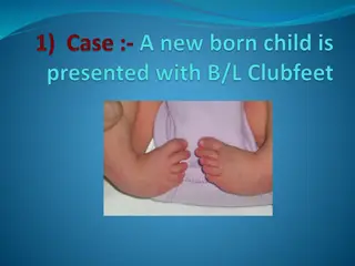
Talipes Equinovarus Common musculoskeletal birth defect that if untreated causes long-term functional disability Varying severity Photos courtesy of CDC-Beijing Medical University collaborative project Talipes | 6
Differential Diagnosis Positional clubfoot Can be manipulated into a normal position Most times do not require treatment Clubfoot associated with neuromuscular diagnoses or syndromes, such as spina bifida (sequence) arthrogryposis multiplex congenita congenital myotonic dystrophy diastrophic dysplasia Tibial reduction defect chromosomal abnormalities Tibial and other limb deficiencies can mimic clubfoot Photo courtesy of CDC-Beijing Medical University collaborative project Talipes 7 | 7
Etiology Poorly understood Generally considered to be multifactorial Evidence for a genetic contribution* Prevalence varies among ethnic groups Twin studies showing higher concordance in monozygotic than in dizygotic twins Runs in families Evidence for environmental contribution Smoking *Source: Miedzybrodzka Z. 2003. J. Anat. 202:37 42 Talipes | 8
Diagnosis Talipes equinovarus is usually diagnosed by physical exam, but at times an x- ray may be necessary Clinical evaluation X-Rays Prenatal diagnoses should not be included in surveillance data unless it is confirmed postnatally. Photo courtesy of CDC-Beijing Medical University collaborative project Talipes | 9
Epidemiologic Features of Talipes Equinovarus Feature Typical Findings Varies among ethnic groups Asian populations: 6 per 10,000 live births Caucasians: 10 to 30 per 10,000 live births Polynesian ancestry: 60 per 10,000 live births Prevalence* Sex differences* Male to female ratio 2:1 Bilateral talipes: about 50% of cases Unilateral talipes: right more common than left Laterality* Most common associated conditions: spina bifida (4.4% of children with talipes); cerebral palsy (1.9%), and arthrogryposis (0.9%) Associations** *Source: Parker, et al, Birth Defects Research (Part A) 2009 **Source: Ching et al, Am J Hum Genet 1969. Talipes | 10
Elements for Description Describe laterality: right, left, or bilateral Describe mobility of foot rigid vs flexible Describe additional malformations when present A photograph should be taken; it can be useful for review, but it is not sufficient for confirmation Talipes | 11
Relevant ICD-10 Codes Q66 Congenital deformities of feet (avoid using this general code if more specific information is available) Q66.0 Talipes equinovarus Q66.1 Talipes calcaneovarus Q66.4 Talipes calcaneovalgus Q66.8 Other congenital deformities of feet Clubfoot unspecified Exclusions Clubfoot, positional Clubfoot associated with: - neuromuscular diagnoses (e.g., spina bifida sequence) - syndromes, such as arthrogryposis ,multiplex congenita, congenital myotonic dystrophy, or diastrophic dysplasia Talipes | 12
Talipes Calcaneovarus Excessive dorsiflexion of the foot that allows its dorsum to come into contact with the anterior aspect of the lower leg the toes point upward, the arch is flat Talipes | 13
Talipes Calcaneovalgus The axis of calcaneovalgus deformity is in the tibiotalar joint, where the foot is positioned in extreme hyperextension. The foot has an "up and out" appearance, with the dorsal forefoot practically touching the anterior aspect of the ankle and lower leg. Illustration source: http://www.human-phenotype-ontology.org/hpoweb/showterm?id=HP:0001884 Talipes | 14
Questions? If you have questions please send an email to centre@icbdsr.org or to birthdefectscount@cdc.gov Talipes | 15


