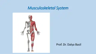
Important Functions of Bones in the Human Body
The human skeleton serves crucial roles in supporting and protecting vital organs, generating blood cells, storing minerals like calcium, facilitating muscle attachments, and enabling body movement. Bones come in various shapes and structures, encompassing features like periosteum, epiphysis, diaphysis, medullary cavity, and compact bone organization. Understanding bone development, maintenance, and repair processes helps comprehend skeletal health and fracture management.

Download Presentation

Please find below an Image/Link to download the presentation.
The content on the website is provided AS IS for your information and personal use only. It may not be sold, licensed, or shared on other websites without obtaining consent from the author.If you encounter any issues during the download, it is possible that the publisher has removed the file from their server.
You are allowed to download the files provided on this website for personal or commercial use, subject to the condition that they are used lawfully. All files are the property of their respective owners.
The content on the website is provided AS IS for your information and personal use only. It may not be sold, licensed, or shared on other websites without obtaining consent from the author.
E N D
Presentation Transcript
The 2ndcorss L1 Senior Anesthesia Technologist BCS. Anesthesia. and IC diploma. Community health Muneer Salman Hasan Senior Anesthesia Technologist BCS. Anesthesia. and IC diploma. Community health KarrarNader AL-Taie
Cardiorespiratory Arrest Step 1: Early Recognition of Sudden Cardiac Arrest Check Responsiveness Check response: Gently tap the patient on his/her shoulders and check for a response Step 2: Activate the Emergency System If Patient Is Unresponsive Activate the emergency system if present in your hospital or just shout for help. Get the defibrillator or send someone to get it. Step 3: Check the Breathing and Pulse Check breathing no breathing or no normal breathing (i.e., only gasping). Check pulse Healthcare Providers should check the pulse simultaneously while checking for breathing, to minimize delay in detection of cardiac arrest and initiation of CPR. Check the pulse using a central pulse (carotid or femoral) for no more than 10 s. If pulse is not felt in 10 s or there is any doubt, start chest compression. Lay rescuers need not check for a pulse.
Step 4: Start Cardiopulmonary Resuscitation (CPR): Initiate Chest Compressions Before Giving Rescue Breaths (Airway, Breathing, and Circulation [ABC] Is Now Circulation, Airway, and Breathing [CAB]) Positioning The victim should lie supine on a hard surface. The rescuer should kneel beside the victim s thorax (either side). Keep arms straight, elbows locked, and the shoulder and the hands of the provider should be in vertical line. Hand placement: Place the heel of the hand on the lower half of the victim s sternum in the center (middle) of the chest, between the nipples, and then place the heel of the second hand on top of the first so that the hands are overlapped and parallel. Interlock fingers to avoid compression on the ribs.
Technique During CPR, remember to push hard and push fast___(all you need is two hands): 1- Compressions rate 100 120/min. 2- Depth of sternal compression At least 2 inches i.e. 5 cm (at least one-third anteroposteriordiameter in children and infants). Avoid excessive chest compression depths (greater than 6 cm). 3- Compression ventilation ratio the adult patient 30:2 (with one or more rescuers) and in the child or infant 30:2 (with one rescuer) and 15:2 (with more than one rescuer). 4- Compression relaxation ratio 1:1 (allow complete recoil of the chest). 5- Perform five cycles (approximately 2 min) of compression and ventilation (ratio 30:2). 8- Give 2 min of uninterrupted CPR. Limit any interruption
Ventilation Untrained lay rescuers should not attempt ventilation and should provide Compression-only CPR . 30 compressions to 2 breaths. After every 30 chest compressions, two slow rescue breaths (each breath over 1 s) can be given using the face mask and the AMBU bag (with a reservoir bag) to deliver 100% oxygen. Before you start ventilation, open the airway using a jaw thrust or a head tilt/chin lift maneuver. If cervical spine injury is suspected, airway should be opened using a jaw thrust without head extension (head tilt/chin lift)). Give one breath over 1 s. Rapid ventilation should be avoided as it can lead to gastric insufflations with increase in the risk of aspiration. Give sufficient tidal volume to ensure visible chest rise. Reposition mask if there is a leak and insert an oral or nasal airway if there is airway obstruction due to tongue fall. The use of cricoid pressure during ventilation is generally not recommended.
Step 5: Attach the Defibrillator (Automated External Defibrillators [AED] or Manual Defibrillators) and Shock If Indicated As soon as an AED/manual defibrillator is available, attach it, check rhythm and deliver shock if indicated, i.e., in ventricular fibrillation (VF) and pulseless ventricular tachycardia (VT). Always prefer a biphasic defibrillator; if biphasic defibrillator is unavailable, monophonic defibrillator can be used. Ensure that no one is touching the patient before you deliver the shock. Shock energy (a)Biphasic: Use the manufacturer s recommendation (120 200 J); if unknown, use maximum available. Second and subsequent doses should be equivalent, and higher doses may be considered if available. (b) Monophasic: 360 J (c)(in children and infants, use 2 4 J/Kg first and 4 J/Kg for subsequent shocks; higher energy may be considered but not to exceed 10 J/Kg).
Technique No pulse check is recommended after defibrillation; resume CPR immediately. Reattach the defibrillator after every 2 min of CPR. Reduce time between the last compression and shock delivery There is no upper limit to the number of shocks you give.. AEDs can now be used even in infants with a pediatric Electric pacing is not recommended for routine use in cardiac arrest. If it is not a shockablerhythm, resume CPR for 2 min and then re attach the defibrillator
Advanced Cardiac Life Support Step 6: Drug Therapy Use intravenous (IV) or intraosseous (IO) route for bolus delivery of drugs. For IV use, give the bolus drug followed by a 20-mL saline push and raise the extremity. If both IV and IO are unavailable, then tracheal route may be used. Epinephrine, naloxone and lidocaine. (Use 2 2 times the dose diluted in 5 10 mL of distilled water or saline for the tracheal route) Give a vasopressor soon after giving the shock. Epinephrine IV/IO dose should be 1 mg every 3 5 min. Amiodaroneshould be given when VF/VT is unresponsive to CPR, defibrillation, and vasopressor therapy. IV/IO first dose should be 300 mg bolus, and the second dose should be 150 mg (after 3 5 min if VF/VT recurs or persists). This may be followed by a 24-h infusion. (Use lidocaineonly if amiodarone is unavailable.) Atropine should not be used during pulseless electrical activity or asystole
Step 7: Advanced Airway the endotracheal tube or the supraglottic airway (laryngeal mask airway, esophageal tracheal tube Combitube, or laryngeal tube). Continue bag mask ventilation if advanced airway is not placed. Confirm the placement of advanced airway by the clinical method (chest expansion and breath sound), and in addition, use the waveform capnography Once advanced airway is in place, give 10 breaths per minute
Step 8: Treat Reversible Causes During each 2-min period of CPR, review the most frequent causes five H s and five T s to identify factors that may have caused the arrest or may be complicating the resuscitation. Step 9: Monitor the CPR Quality Throughout Resuscitation delivering high-quality CPR. giving compressions of adequate rate and depth allowing complete chest recoil between compressions minimizing interruptions in compressions avoiding excessive ventilation, rotating the compressor every 2 min. waveform capnography to monitor end tidal CO2 if available If intra-arterial pressure monitoring is already present and the diastolic pressure is less than 20 mmHg, attempt should be made to improve quality of CPR.
Step 10: Return of Spontaneous Circulation ROSC Return of spontaneous circulation (ROSC) can be confirmed by return of pulse or blood pressure or abrupt sustained increase in PetCO2 Step 11: PostcardiacArrest Care After ROSC The goal should be to optimize cardiopulmonary function and vital organ perfusion. Transfer the patient to an appropriate hospital or ICU with facility to deliver postcardiacarrest care. Optimize ventilation to minimize the lung injury. Do chest X-ray to confirm airway position and to diagnose pneumonia/pulmonary edema. Use lung-protective ventilation if there is pulmonary dysfunction; adjust settings using blood gas values. Avoid excessive ventilation and hyperoxia


: Initiate")