Improved Early Detection of Hepatocellular Carcinoma with Biomarker Screening Algorithms
Discover the importance of early detection in hepatocellular carcinoma (HCC) and the challenges with current surveillance practices. Learn about longitudinal AFP screening algorithms and insights from the HALT-C trial for advanced fibrosis or cirrhosis. Stay informed on advancements in HCC screening to improve patient outcomes.
Download Presentation

Please find below an Image/Link to download the presentation.
The content on the website is provided AS IS for your information and personal use only. It may not be sold, licensed, or shared on other websites without obtaining consent from the author. If you encounter any issues during the download, it is possible that the publisher has removed the file from their server.
You are allowed to download the files provided on this website for personal or commercial use, subject to the condition that they are used lawfully. All files are the property of their respective owners.
The content on the website is provided AS IS for your information and personal use only. It may not be sold, licensed, or shared on other websites without obtaining consent from the author.
E N D
Presentation Transcript
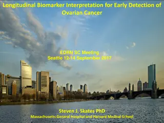
Improved early detection of hepatocellular carcinoma with longitudinal biomarker screening algorithms Nabihah Tayob The University of Texas MD Anderson Cancer Center EDRN Steering Committee Meeting, Seattle September 12-14, 2017 Nabihah Tayob, MD Anderson Cancer Center HCC Screening Algorithms
Early Detection of Hepatocellular Carcinoma? Hepatocellular carcinoma (HCC) has five-year survival <12%. Early (or very early) stage HCC: Surgical resection, liver transplantation and loco-regional therapies. 5-year survival is 47-84% Later stage HCC (symptomatic): 5-year survival is <10% Key to reducing HCC mortality: early detection. Nabihah Tayob, MD Anderson Cancer Center HCC Screening Algorithms
Current Surveillance Practice Key risk factor is cirrhosis: accounts for 80-90% of HCC cases. Hepatitis B and C infection Alcoholic liver disease Non-alcoholic fatty liver disease AASLD guidelines: screening every 6 months via Ultrasonography mandatory -Fetoprotein (AFP) optional but common Positive screen CT or MRI Nabihah Tayob, MD Anderson Cancer Center HCC Screening Algorithms
Current Surveillance Practice Problems with ultrasonography: Operator dependent Difficult to perform in obese patients Sensitivity for early stage HCC in clinical practice: 32% Serum AFP is a diagnostic biomarker that is widely used in screening. Sensitivity 41-100% Specificity 70-90% Nabihah Tayob, MD Anderson Cancer Center HCC Screening Algorithms
Incorporating longitudinal AFP Lee et al (2013): standard deviation and rate of increase of AFP vs current AFP. White et al (2015): current AFP and rate of increase in AFP within the last year vs current AFP. McIntosh and Urban (2003): parametric empirical Bayes (PEB) approach. Skates, Pauler and Jacobs (2001): posterior risk from Bayesian hierarchical changepoint model. Nabihah Tayob, MD Anderson Cancer Center HCC Screening Algorithms
HALT-C Trial Nabihah Tayob, MD Anderson Cancer Center HCC Screening Algorithms
HALT-C trial design Enrollment criteria: Chronic hepatitis C. Stage 3 fibrosis (Advanced fibrosis or cirrhosis). Radiological imaging to exclude HCC. History of non-response to interferon-based therapy. Randomization: long-term low-dose pegylated interferon therapy v.s. no treatment Follow-up schedule: Nabihah Tayob, MD Anderson Cancer Center HCC Screening Algorithms
HALT-C Trial Randomized pa,ents (N=1050) Missing HCC status (N=2) Randomized pa,ents with complete clinical data (N=1048) Advanced fibrosis at baseline biopsy (N=621) Cirrhosis at baseline biopsy (N=427) No HCC, <12m follow- up (N=23) No HCC, >12m follow- up (N=558) HCC confirmed (N=40) No HCC, <12m follow- up (N=18) No HCC, >12m follow- up (N=361) HCC confirmed (N=48) HCC diagnosis: histology or imaging (with or without AFP). All diagnoses adjudicated by a panel of investigators. Nabihah Tayob, MD Anderson Cancer Center HCC Screening Algorithms
Parametric Empirical Bayes (PEB) Method: Has a deviation in the biomarker trajectory for a particular patient occurred? Specify a model for the biomarker in control patients. End of follow-up Within-subject variability P2 Between- subject variability Popula8on Mean AFP P1 Parameters are estimated using control patients only. Nabihah Tayob, MD Anderson Cancer Center HCC Screening Algorithms
PEB threshold: Suppose a patient has completed n screenings. Compare current AFP to a individually tailored threshold for that patient. Estimate mean AFP level for patient: PEB estimate = Weight 1 population mean + Weight 2 sample mean PEB Rule: Yn+ 1> PEB estimate+c1 SD of PEB estimate Standard Threshold (ST) Rule: Yn+ 1> c2 Select c1and c2to control the false positive rate. Nabihah Tayob, MD Anderson Cancer Center HCC Screening Algorithms
PEB threshold: Suppose a patient has completed n screenings. Compare current AFP to a individually tailored threshold for that patient. Estimate mean AFP level for patient: PEB estimate = Weight 1 population mean + Weight 2 sample mean PEB Rule: Yn+ 1> PEB estimate+c1 SD of PEB estimate Standard Threshold (ST) Rule: Yn+ 1> c2 Select c1and c2to control the false positive rate. Nabihah Tayob, MD Anderson Cancer Center HCC Screening Algorithms
8 6 log(AFP+0.01) 4 2 0 4 3 2 1 Years to diagnosis Nabihah Tayob, MD Anderson Cancer Center HCC Screening Algorithms
8 6 log(AFP+0.01) 4 2 0 4 3 2 1 Years to diagnosis Nabihah Tayob, MD Anderson Cancer Center HCC Screening Algorithms
8 6 log(AFP+0.01) 4 2 0 4 3 2 1 Years to diagnosis Nabihah Tayob, MD Anderson Cancer Center HCC Screening Algorithms
8 6 log(AFP+0.01) 4 2 0 4 3 2 1 Years to diagnosis Nabihah Tayob, MD Anderson Cancer Center HCC Screening Algorithms
8 6 log(AFP+0.01) 4 2 0 4 3 2 1 Years to diagnosis Nabihah Tayob, MD Anderson Cancer Center HCC Screening Algorithms
8 6 log(AFP+0.01) 4 2 0 4 3 2 1 Years to diagnosis Nabihah Tayob, MD Anderson Cancer Center HCC Screening Algorithms
8 6 log(AFP+0.01) 4 2 0 4 3 2 1 Years to diagnosis Nabihah Tayob, MD Anderson Cancer Center HCC Screening Algorithms
8 6 log(AFP+0.01) 4 2 0 4 3 2 1 Years to diagnosis Nabihah Tayob, MD Anderson Cancer Center HCC Screening Algorithms
8 6 log(AFP+0.01) 4 2 0 4 3 2 1 Years to diagnosis Nabihah Tayob, MD Anderson Cancer Center HCC Screening Algorithms
8 6 log(AFP+0.01) 4 2 0 4 3 2 1 Years to diagnosis Nabihah Tayob, MD Anderson Cancer Center HCC Screening Algorithms
8 6 log(AFP+0.01) 4 2 0 4 3 2 1 Years to diagnosis Nabihah Tayob, MD Anderson Cancer Center HCC Screening Algorithms
8 6 log(AFP+0.01) 4 2 0 4 3 2 1 Years to diagnosis Nabihah Tayob, MD Anderson Cancer Center HCC Screening Algorithms
8 6 log(AFP+0.01) 4 2 0 4 3 2 1 Years to diagnosis Nabihah Tayob, MD Anderson Cancer Center HCC Screening Algorithms
8 6 log(AFP+0.01) 4 2 0 4 3 2 1 Years to diagnosis Nabihah Tayob, MD Anderson Cancer Center HCC Screening Algorithms
8 6 log(AFP+0.01) 4 2 0 4 3 2 1 Years to diagnosis Nabihah Tayob, MD Anderson Cancer Center HCC Screening Algorithms
Evaluating the screening algorithms False positive rate (FPR): proportion of positive screens among all the screenings conducted in the control group. Specificity=1-FPR. True positive rate (TPR)/Sensitivity: proportion of HCC cases with at least one positive screening during the pre-diagnostic period. Nabihah Tayob, MD Anderson Cancer Center HCC Screening Algorithms
ROC results: cirrhosis at baseline 1.0 0.8 True Positive Rate (Patients) 0.6 0.4 PEB, AUC=0.93 0.2 ST, AUC=0.84 0.0 0.0 0.2 0.4 0.6 0.8 1.0 False Positive Rate (Screens) Bootstrap p-value value<0.0005. Tayob N, Lok ASF, Do K, Feng Z. Improved Detection of Hepatocellular Carcinoma by Using a Longitudinal Alpha-Fetoprotein Screening Algorithm. Clin Gastroenterol Hepatol. 2016;14(3):469-475.e2 Nabihah Tayob, MD Anderson Cancer Center HCC Screening Algorithms
ROC results: cirrhosis at baseline 1.0 0.8 True Positive Rate (Patients) 0.6 0.4 PEB 0.2 ST 0.0 0.00 0.05 0.10 0.15 0.20 False Positive Rate (Screens) Nabihah Tayob, MD Anderson Cancer Center HCC Screening Algorithms
Summary: PEB method Incorporating AFP screening history patient specific thresholds higher sensitivity AFP does not increase in all HCC cases. Incorporate additional biomarkers into a longitudinal screening algorithm to identify all the disease subtypes. Nabihah Tayob, MD Anderson Cancer Center HCC Screening Algorithms
Incorporating longitudinal DCP Des-gamma carboxy-prothrombin (DCP) is a blood-based biomarker that is part of the FDA approved triplicate. Previous generation assay for lectin-bound AFP (AFP-L3%) only. In the HALT-C Trial: Tayob N, Stingo F, Do K-A, Lok ASF, Feng Z. A Bayesian screening approach for hepatocellular carcinoma using multiple longitudinal biomarkers. Biometrics. Nabihah Tayob, MD Anderson Cancer Center HCC Screening Algorithms
Bayesian screening algorithm Our goals: Exploit all the available information in multiple longitudinal biomarkers. Robust and computationally efficient. Unevenly spaced biomarker measures. Missing data. Nabihah Tayob, MD Anderson Cancer Center HCC Screening Algorithms
Decision rule: posterior risk Pr(HCC|All Data) Pr(no HCC|All Data) Pr(AllData|HCC) Pr(AllData|no HCC) Posterior Risk = Pr(HCC) = Pr(no HCC) Specify a joint model for biomarkers in cases and controls. Prior probability of disease: target population for surveillance. Nabihah Tayob, MD Anderson Cancer Center HCC Screening Algorithms
Decision rule: posterior risk Pr(HCC|All Data) Pr(no HCC|All Data) Pr(AllData|HCC) Pr(AllData|no HCC) Posterior Risk = Pr(HCC) = Pr(no HCC) Specify a joint model for biomarkers in cases and controls. Prior probability of disease: target population for surveillance. Nabihah Tayob, MD Anderson Cancer Center HCC Screening Algorithms
Decision rule: posterior risk Pr(HCC|All Data) Pr(no HCC|All Data) Pr(AllData|HCC) Pr(AllData|no HCC) Posterior Risk = Pr(HCC) = Pr(no HCC) Specify a joint model for biomarkers in cases and controls. Prior probability of disease: target population for surveillance. Nabihah Tayob, MD Anderson Cancer Center HCC Screening Algorithms
Hierarchical model for biomarker level in controls Yijk End of follow- u p ik tij di Nabihah Tayob, MD Anderson Cancer Center HCC Screening Algorithms
Hierarchical model for biomarker level in cases We define an unobserved Iikto distinguish between two possible models for the kthmarker in cases. If Iik= 0 Yijk Clinical Diagnosis ik tij di Nabihah Tayob, MD Anderson Cancer Center HCC Screening Algorithms
Hierarchical model for biomarker level in cases If Iik= 1 Yijk Clinical Diagnosis ik ik tij ik di Nabihah Tayob, MD Anderson Cancer Center HCC Screening Algorithms
Connecting the biomarker models The indicator variables follow a Markov Random Field distribution. What does this mean? The probability that we have observed a changepoint in one biomarker depends on whether we have observed changepoints in other biomarkers. This allows us to borrow information across the biomarkers to detect changepoints. Nabihah Tayob, MD Anderson Cancer Center HCC Screening Algorithms
Calculating the posterior risk A Markov chain Monte Carlo (MCMC) procedure is constructed to sample from the joint posterior distribution for all the model parameters. Gibbs sampler when full conditionals are easily computed. Metropolis-Hasting algorithm when full conditionals are not straightforward. Reversible jump procedure (Green, 1995) when the dimension of the parameter space depends on Iik. Monte Carlo integration is used to get estimates of posterior predictive probabilities. Calculate posterior risk at each screening visit Nabihah Tayob, MD Anderson Cancer Center HCC Screening Algorithms
Results from HALT-C Trial Apply screening algorithm with log(AFP) and log(DCP + 1) in cirrhosis patients. Ideal study: Training dataset: obtain samples from joint posterior distribution for all the model parameters. Testing dataset: Estimate the posterior risk in each patient at each screening visit and evaluate performance of the proposed screening algorithm. Use 10-fold cross validation to evaluate the proposed screening algorithm. Nabihah Tayob, MD Anderson Cancer Center HCC Screening Algorithms
Screening algorithms: mFB: multivariate fully Bayesian screening algorithm uFB: univariate fully Bayesian screening algorithm (Skates et al, 2001) uEB: univariate parametric empirical Bayesian screening algorithm (McIntosh and Urban, 2003) Nabihah Tayob, MD Anderson Cancer Center HCC Screening Algorithms
ROC results 1.0 mFB 0.8 True Positive Rate (Patients) uEB AFP 0.6 uFB DCP 0.4 uEB DCP uFB AFP 0.2 0.00 0.05 0.10 0.15 0.20 False Positive Rate (Screens) Nabihah Tayob, MD Anderson Cancer Center HCC Screening Algorithms
Early detection of HCC Compare the time of the first positive screen for each method: During the entire screening period T=1 years Nabihah Tayob, MD Anderson Cancer Center HCC Screening Algorithms
Entire screening period uEB Biomarker mFB-J uFB mFB J first other method first same time 77.00% 60.50% log(AFP) log(DCP+1) 81.00% 70.50% 89.50% 68.00 52.00 50.50 47.00 44.50 36.50 33.00 29.00 26.50 25.00 24.50 20.00 19.00 13.00 11.50 uFB AFP uFB DCP uEB AFP uEB DCP mFB I mFB positive before the uEB with AFP 29% of the time. uEB with AFP positive before mFB 26.5% of the time. Nabihah Tayob, MD Anderson Cancer Center HCC Screening Algorithms
One year prior to clinical diagnosis uEB Biomarker mFB-J uFB 91.50 mFB J first other method first 83.50 same time 57.00% 56.48% log(AFP) log(DCP+1) 53.00% 43.52% 69.00 59.50% 50.93 44.44 35.18 29.63 25.93 18.50 13.89 12.50 10.50 6.00 4.50 4.00 uFB AFP uFB DCP uEB AFP uEB DCP mFB I mFB positive before the uEB with AFP 12.5% of the time. uEB with AFP positive before mFB 18.5% of the time. Nabihah Tayob, MD Anderson Cancer Center HCC Screening Algorithms
Applying the PEB algorithm in a VA HCV cohort HALT-C is a randomized clinical trial with 3 month surveillance windows. AFP measured at local laboratories for each University hospital. How does PEB algorithm perform in a regular clinical setting? Department of Veterans Affairs (VA) is the largest integrated health-care provider in the United States. Important setting to study the PEB algorithm for AFP. Nabihah Tayob, MD Anderson Cancer Center HCC Screening Algorithms
VA HCV cohort: 12,124 patients with cirrhosis diagnosed between 1997 and 2005 and at least one AFP test. 11,222 with no HCC ever 902 with HCC eventually Use IDC-9 diagnostic codes to identify patients with cirrhosis. HCC diagnosis first identified with IDC-9 codes and confirmed with manual review. AFP pulled from the laboratory records at the VA. Nabihah Tayob, MD Anderson Cancer Center HCC Screening Algorithms
Results: HALT-C vs VA Table : Patient-level true positive rate (TPR) for hepatocellular carcinoma corresponding to 10% screening-level false positive rate. HALT-C 60.4 77.1 VA 53.9 63.6 ST PEB HALT-C: 17% increase in TPR when prior AFP is incorporated. VA: 10% increase in TPR when prior AFP is incorporated. Nabihah Tayob, MD Anderson Cancer Center HCC Screening Algorithms
Results: HALT-C vs VA Table : Patient-level true positive rate (TPR) for hepatocellular carcinoma corresponding to 10% screening-level false positive rate. HALT-C 60.4 77.1 VA 53.9 63.6 ST PEB HALT-C: 17% increase in TPR when prior AFP is incorporated. VA: 10% increase in TPR when prior AFP is incorporated. Nabihah Tayob, MD Anderson Cancer Center HCC Screening Algorithms

