Incidental Findings in Cone Beam Computed Tomography Scans: A Review
Review the prevalence and risk of incidental findings in CBCT scans, analyzing advantages, disadvantages, methods, and inclusion criteria. Explore the impact on dentistry practice.

Download Presentation

Please find below an Image/Link to download the presentation.
The content on the website is provided AS IS for your information and personal use only. It may not be sold, licensed, or shared on other websites without obtaining consent from the author.If you encounter any issues during the download, it is possible that the publisher has removed the file from their server.
You are allowed to download the files provided on this website for personal or commercial use, subject to the condition that they are used lawfully. All files are the property of their respective owners.
The content on the website is provided AS IS for your information and personal use only. It may not be sold, licensed, or shared on other websites without obtaining consent from the author.
E N D
Presentation Transcript
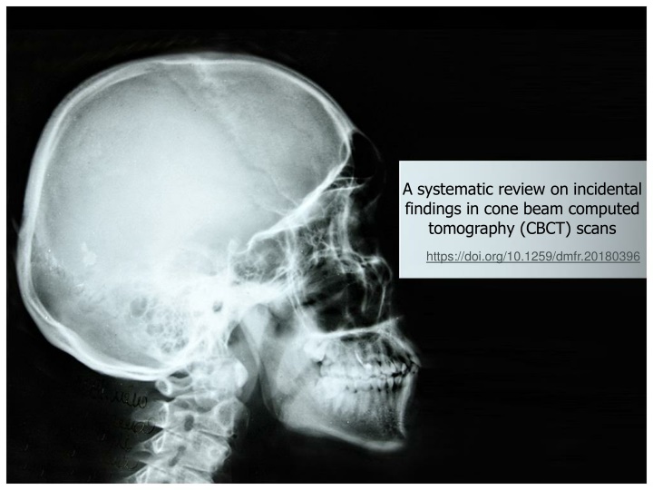
A systematic review on incidental findings in cone beam computed tomography (CBCT) scans https://doi.org/10.1259/dmfr.20180396
CBCT Advantages: Proper resolution for Large structures Intricate tooth anatomy Multiplanar imaging 2D & 3D images different perspective Different FOV Large FOV: Maxilla, mandible, sinus, airway, cranial structures Disadvantages: Lack of soft tissue window Higher image noise Higher radiation
Incidental findings Any occult finding discovered upon assessing radiographic image taken for other purposes High prevalence in CBCT scans Aim of this study evaluation of the frequency and risk of IFs using CBCT
PRISMA Statement Search Criteria Databases PubMed, Embase (through Ovid), MEDLINE (through Ovid),Web of Science, and Scopus Primary keywords cone-beam computed tomography incidental finding Risk of Bias: STROBE guidelines
Phase 1 Inclusion criteria Human participants of all ages IFs of head & neck area English peer-reviewed articles Exclusion criteria Case reports & case series Craniofacial syndrome Studies focusing on one type of IF
Phase 2 Inclusion criteria Studies focusing on one field of dentistry Exclusion criteria Studies not presenting a dental focus Systematic reviews
Methods and materials Phase 2 Inclusion criteria Studies focusing on one field of dentistry Exclusion criteria Studies not presenting a dental focus Systematic reviews
Methods and materials Table 2
Price et al TMJ findings more commonly found in older age Pette et al Vertebral pathological findings 28.67 times more in 65 years old participants Mutalik CBCT scan for dental implants is in tandem with an increase in an increase in age and IFs Damaskos IFs (e.g.: calcification of atherosclerotic plaques) increases with age Pliska et al & Drage et al Younger patients IFs: tooth impaction IFs: Highest frequency: mucous retention cyst (55.1%) Lowest frequency: malignancies (1.4%)
Calcification of Atherosclerotic Plaque Carotid Fat deposit accumulation slowing down the blood of brain stroke Frequency 3 of 10 studies (2-11.6%) Allareddy et al: 57/1000 Pette et al: 37/318 Price et al: 17/300 The other 7 study? Cutting off the carotid space from FOV Not considering CAC as an IF
Calcification of Atherosclerotic Plaque Damaskos et al (ACs): Intracranial: 161 of 484 (33.26%) Extracranial: 150 of 484 (30.99%) More frequent in male Increased with age
Pathologies & Malignancies At least 2 of 10 articles: Dentigenerous cyst: 0.6- 2.6% Ameloblastoma: 0.2- 0.8% OKC: 0.2- 0.5% Nasopalatine/ incisive canal cyst: 0.3-2.5% Giant cell lesions: 0.1-0.5% Allareddy et al (large sample size) Residual cyst (0.7%), Osteosclerosis (0.5%), cementoblastoma(0.4%), osteomyelitis (0.2%), radioosteonectosis (0.1%), chemoosteonecrosis (0.1%), odontoma (0.4%), periapical cyst (1.78%)
Pathologies & Malignancies Total frequency of malignancy: 0.3%(Allareddy et al)- 1.4% (Warhekar et al) Significant & critical to identify Allareddy et al s malignancies: In sella region damaging sella turcica Metastatic lesions in breast & prostate Warhekar et al : Orofacial malignancies The importance of evaluation of CBCT scan beyond maxillomandibular regions
Non-threatening IFs Sinusitis 41.7%, 7 of 10 studies Endodontic lesions 11.8% Untreated Bone loss, tooth loss, periodontal disease Loss of adjacent cortical plates, perforation of inferior alveolar canal Also can be evaluate by conventional radiographic tools
CBCT Characteristics & Disadvantages Higher radiation Small FOV: 11-652 Sv Large FOV: 30-1073 Sv Conventional: <8.3 Sv Panoramic: 2.7-23 Sv Maxillomandibular CT: 180-2100 Sv Lack of soft tissue window Higher image noise Higher cost Main disadvantage: Dentists are accustomed to small FOV
CBCT Characteristics & Disadvantages Primary evaluation tool: Conventional radiographs Secondary evaluation tool: CBCT scan
Limitations Specific focus on a specialization of dentistry IFs not confirmed by lab results or biopsy Significant variation among reviewers 4 articles: 1 OMFR 4 articles: 2 independent OMFRs 1 article: not specified 1 article: 13 masked independent OMFRs Different CBCT machines, FOV, imaging parameters 4 of 10 articles explained the term of incidental finding
Similar Articles Mutalik et al Possible missing of IFs due to: Large FOV & unfamiliarity of dentists for associated anatomical areas Solution: a holistic view by dentists & radiologists (first responders to critical findings)
Similar Articles Edwards et al (2014) Same 5 articles IF frequency: Edwards: 24.6- 93.4% This article: 24.6- 94.3% Additionally 5 articles: published after 2012 increase in CBCT use more focus on IFs Same tools assessing the methodological quality Edwards: moderate to low in bias(60-93%) This article: moderate to low in bias(44-94%)
Importance of CBCT complete readings & diagnosis of IFs Risk of potential bias of all included studies due to the research design Primary evaluation tool: Conventional radiographs Secondary evaluation tool: CBCT scan The importance of evaluation of CBCT scan beyond maxillomandibular regions