Infectious Footrot and Leptospirosis in Sheep and Other Livestock
Infectious footrot in sheep is caused by Dichelobacter nodosus and can vary in severity. Leptospirosis affects multiple livestock species and is caused by Leptospira interrogans. Both diseases have distinct etiology, clinical findings, and treatment options. Understanding the epidemiology, pathogenesis, and clinical pathology is essential for effective diagnosis and control measures. Explore more about these infectious diseases prevalent in warm, wet climates.

Download Presentation

Please find below an Image/Link to download the presentation.
The content on the website is provided AS IS for your information and personal use only. It may not be sold, licensed, or shared on other websites without obtaining consent from the author.If you encounter any issues during the download, it is possible that the publisher has removed the file from their server.
You are allowed to download the files provided on this website for personal or commercial use, subject to the condition that they are used lawfully. All files are the property of their respective owners.
The content on the website is provided AS IS for your information and personal use only. It may not be sold, licensed, or shared on other websites without obtaining consent from the author.
E N D
Presentation Transcript
INFECTIOUS FOOTROT IN SHEEP ETIOLOGY Dichelobacter nodosus. Strains vary in virulence to produce benign and virulent footrot. EPIDEMIOLOGY Main source of infection is lesion discharge from other infected sheep. D. nodosus typically survives in the environment for only a few days. Highlycontagious disease with high attack rate in warm, wet conditions. Lesions are present on both claws of the foot and commonly in more than one foot. Significant effect on productivity. PATHOGENESIS Maceration of the interdigital skin from prolonged wet conditions underfoot allow for infection with F. necrophorum. This initial local dermatitis associated with infection with F. necrophorum at the skin and the skin horn junction may progress no further, but the hyperkeratosis induced by this infection facilitates infection by D. nodosus if it is present. The preliminary dermatitis has beennamed ovine interdigital dermatitis and is also called foot scald.
CLINICAL FINDINGS Inflammation of the skin at the skin horn junction in the interdigital area with underrunning of the soft horn in benign (nonprogressive) footrot. Progressesto underrunning of the hard horn and inflammation of the sensitive laminae invirulent (progressive) footrot and severe lameness. CLINICAL PATHOLOGY Gram-stained smears and culture to confirm the presence of the organism; protease and polymerase chain reaction (PCR) tests for strain virulence. TREATMENT AND CONTROL Topical treatment with bactericides in footbaths at time of transmission to minimize new infections, parenteral antibiotics for treating virulent footrot, vaccination, culling. Goats and cattle can carry D. nododus and so must be included in control programs.
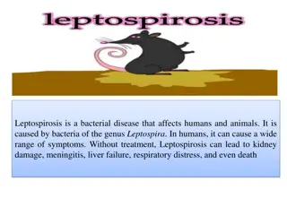
LEPTOSPIROSIS Aetiology Leptospira interrogans (many distinct serovars) and L. borgpetersenii (many distinct serovars) are found. Epidemiology Worldwide distribution, most commonly in warm, wet climates. Occurs in cattle, sheep and goats, pigs, and horses. Host-adapted (maintenance or reservoir) and non host-adapted (accidental or incidental) leptospirosis is dependent on the response of each species to particular serovars. Prevalence of infection is greater than incidence of clinical disease. Transmission by urine of infected animals; some wildlife species may transmit to cattle. Ground surface moisture is the most important factor for persistence of the organism; major zoonosis. Signs Acute, subacute, and chronic forms; there is fever, acute haemolytic anemia, changes in milk, stillbirths, abortion in all species (especially pigs), weak neonates, infertility, milk drop syndrome, and periodic ophthalmia (recurrent uveitis in horse).
Clinical pathology Demonstration and/or culture of organism in blood, urine, cervicovaginal mucus, body fluids, and tissues. Serologic tests, primarily macroscopic agglutination test, and ELISA and DNA probes are done. Lesions Anemia, jaundice, hemoglobinuria, serous hemorrhages, autolysis of aborted fetuses, fetal hepatitis, and nephritis are observed. Diagnostic confirmation Culture or demonstrate organism in body fluids or tissues, and there are high serum titers. Treatment Antimicrobials used to treat acute infection and eliminate leptospiruria. Control Antimicrobials are used to eliminate carriers; vaccination is done with vaccines containing serovars that are causing the disease in that geographic area.