Insight into Giardia lamblia: Morphological Features, Life Cycle, and Clinical Manifestations
Learn about Giardia lamblia, a cosmopolitan flagellate found in the duodenum and upper jejunum. Explore its trophozoite and cyst stages, habitat, life cycle, and pathogenicity causing diarrhea, malabsorption, and other clinical features.

Download Presentation

Please find below an Image/Link to download the presentation.
The content on the website is provided AS IS for your information and personal use only. It may not be sold, licensed, or shared on other websites without obtaining consent from the author. If you encounter any issues during the download, it is possible that the publisher has removed the file from their server.
You are allowed to download the files provided on this website for personal or commercial use, subject to the condition that they are used lawfully. All files are the property of their respective owners.
The content on the website is provided AS IS for your information and personal use only. It may not be sold, licensed, or shared on other websites without obtaining consent from the author.
E N D
Presentation Transcript
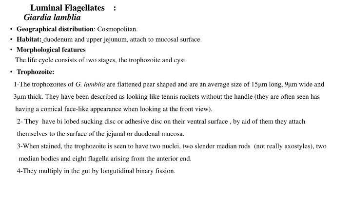
Luminal Flagellates : Giardia lamblia Geographical distribution: Cosmopolitan. Habitat: duodenum and upper jejunum, attach to mucosal surface. Morphological features The life cycle consists of two stages, the trophozoite and cyst. Trophozoite: 1-The trophozoites of G. lamblia are flattened pear shaped and are an average size of 15 m long, 9 m wide and 3 m thick. They have been described as looking like tennis rackets without the handle (they are often seen has having a comical face-like appearance when looking at the front view). 2- They have bi lobed sucking disc or adhesive disc on their ventral surface , by aid of them they attach themselves to the surface of the jejunal or duodenal mucosa. 3-When stained, the trophozoite is seen to have two nuclei, two slender median rods (not really axostyles), two median bodies and eight flagella arising from the anterior end. 4-They multiply in the gut by longutidinal binary fission.
Cyst: It is the infective form of the parasite 1-The cyst is small and oval, measuring 12 m x 8 m and is surrounded by a hyaline cyst wall 2-Its internal structure includes 2 pairs of nuclei grouped at one end. A young cyst contains 1 pair of nuclei. 3-The axostyle with median bodies lies diagnonally, forming a dividing line within cyst wall. 4- Remnants of the flagella and the sucking disc may be seen in the young cyst.
Life Cycle of Giardia lamblia In the small intestine, excystation releases trophozoites (each cyst produces 2 trophozoites) . Trophozoites multiply by longitudinal binary fission remaining in the lumen of the small bowel where they can be free or attached to the mucosa by a ventral sucking disk.
Pathogenicity and Clinical Features 1- Diarrhea with no blood. 2-The trophozoites will cause malabsorption of nutrients by block absorptive activities (villous atrophy or flattening mucosal surfaces) related to fats and fat soluble vitamins (coating). 3-Reduced digestion and absorption may then cause diarrhoea often watery with gas formation, steatorrhoea (greasy stools) will occur. 4-Abdominal pain. 5- Nutritional disturbances. 6-Anorexia, nausea and vomiting. 7-Anemia. 8- Nervous manifestations.
The mechanism of diarrhea in giardiasis The mechanism of diarrhea in giardiasis appears to be multifactorial. The postulated mechanisms are: 1- Coating of intestinal mucosa by a large number of trophozoites, there by interfering with the absorption of fats and other essential nutrients. 2- Damage to the epithelial brush border of the intestinal mucosa 3-Alternations in the motility of the gut, and 4- Increased secretion of fluid into the lumen due to increased adenyl cyclase activity in the enterocyte.
Transmission: transmission by contaminated food and water by sewage, flies or food handler. Laboratory Diagnosis The microscopic identification of Giardia spp. in fecal samples is considered as the gold standard method for the diagnosis of giardiasis. This method is performed to detecting cysts and trophozoites. Because Giardia cysts can be excreted intermittently, multiple stool collections (i.e., three stool specimens collected on separate days) increase test sensitivity. 1-Fecal microscopy examination a- Stool examination by direct wet mount method b- Stool examination after concentration. 2-Stool culture
3-Serological tests a-Serological methods of diagnosis (ELISA)are proving to be useful as means of diagnosis. b- Detection of the trophozoites and cysts can also be achieved by using rapid antigen detection testing kits that specifically detect antigen released by Giardia lamblia. These tests are now commercially available and offer a high degree of sensitivity even on specimens that are preserved and can be detected days before any trophozoites and cysts are shed Treatment For asymptomatic carriers and patients the drug of choice is metronidazole or quinacrine hydrochloride Prevention 1-Asymptomatic reservoirs of infection should be identified & treated. 2-Avoidance of contaminated food and water. 3-Drinking water from lakes and streams should be boiled or filtered. 4-Personal hygiene and health education.