Intestinal Obstruction in Veterinary Medicine
Discover the causes, types, and implications of intestinal obstruction as explained by Dr. Mithilesh Kumar, an esteemed Assistant Professor and Junior Scientist in Veterinary Surgery and Radiology at Bihar Veterinary College. Explore mechanical and functional interferences, site locations, and pathogenesis of obstructions, along with visual illustrations. Learn about a range of conditions such as volvulus, mesenteric torsion, and paralytic ileus that can lead to this serious health issue in animals.
Download Presentation

Please find below an Image/Link to download the presentation.
The content on the website is provided AS IS for your information and personal use only. It may not be sold, licensed, or shared on other websites without obtaining consent from the author. If you encounter any issues during the download, it is possible that the publisher has removed the file from their server.
You are allowed to download the files provided on this website for personal or commercial use, subject to the condition that they are used lawfully. All files are the property of their respective owners.
The content on the website is provided AS IS for your information and personal use only. It may not be sold, licensed, or shared on other websites without obtaining consent from the author.
E N D
Presentation Transcript
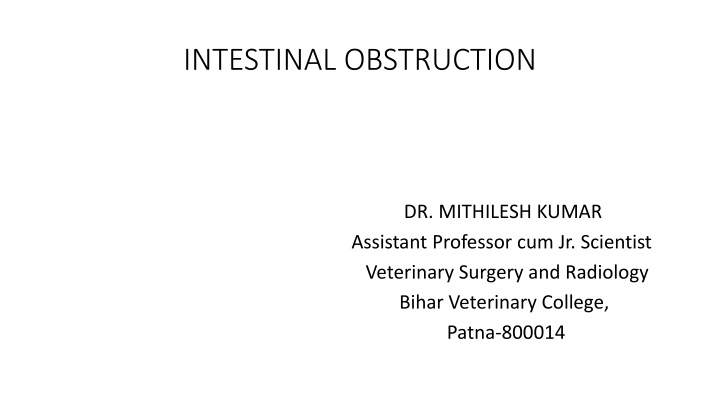
INTESTINAL OBSTRUCTION DR. MITHILESH KUMAR Assistant Professor cum Jr. Scientist Veterinary Surgery and Radiology Bihar Veterinary College, Patna-800014
Mechanical or functional interference cause obstruction. Simple obstruction Strangulated obstruction According to degree of occlusion of lumen Partial Complete
Site of location of obstruction High obstruction Low obstruction AETIOLOGY Mechanical obstruction (i) Intraluminal mass Heavy nodular worm infestation in calves.
Distended small bowel obstructed by bundles of Ascaris lumbricoides worms
(ii) External compression of the intestinal wall Adhesions, fibrous bands, herniation, abscess and neoplasm Trapping of part of intestine GUT TIE:- Castration by traction Vas deferens tears Through gap intestine herniates
VOLVULUS MESENTERIC TORSION INTUSSUSCEPTION PARALYTIC ILEUS Paralysis of a segment or entire length of intestine to produce This is functional obstruction is termed paralytic obstruction
PATHOGENESIS:- Obstruction in flow of ingesta Hypermotility of intestine Stretching of intestine Reduce absorptive capacity Intraluminal pressure increases
Cause venous congestion and fluid loss into intestinal lumen Electrolyte imbalance Change in acid base status In late simple obstruction- Anorexia of intestinal wall Resultant increase in the permeability Fluid loss extensive
Strangulated obstruction- Distensibility of the bowel is magnified due to involvement of circulation Rapid deterioration in animal Hypovolemia Acid base imbalance Abomasal reflex and regurgitation
Strangulated obstruction Veins are occluded Some arterial supply remains intact Extravasation of plasma Rapid multiplication of microbes Release of endotoxins.
Increase in permeability of intestine Absorption of endotoxins and acute fluid loss Endotoxins and hypovolemic shock. Shock is greater than obstruction
More pronounced in proximal obstruction Abomasal reflex. Simple intestinal obstruction- Hypochloraemic hypokalemia metabolic alkalosis Abomasal reflex. The pathogenesis of metabolic alkalosis in ruminants is abomasal reflex
Any abnormality in the movement of gastric juices from abomasum to intestine may produce metabolic alkalosis. Pre-renal azotaemia is observed IO Hemoconcentration- increased PCV.
CLINICAL SIGNS: Acute case blood vessels compromised Pain in initial stage. Buffalo straining Cessation of defecation anorexia Distension of abdomen
In cattle- colic in initial stage Looking towards site of pain Kicking at the abdomen Frequent standing up lying down Paddling of hindlimb.
Respiration rate normal Pulse increased more than 100 beats/minute in cattle. Rectal temp. elevated subnormal in later Faeces not passed Pasty faeces tinged with blood and thick mucus present in rectum. Strangulated obstruction-hypovolemia and endotoximia
Cardiovascular embrassment and depression Condition deteriotes Simple intestinal obstruction live 8-14 days Strangulated Intestinal obstruction live 96 hours. LABORATORATORY EXAMINATION:- PCV increased Azotaemia, hypokalaemia, hypochloraemia and metabolic alkalosis rumen fluid chloride concentration is high
DIAGNOSIS History Clinical signs Rectal examination Laboratory finding Rt flank laparotomy
TREATMENT Rt flank laparotomy Manual exploration of the peritoneal cavity Intraluminal mass found Enterotomy
Minimise and control contamination Affected segment isolated and packed off Inserting 14 gauge needle attached with a long tube Intestinal clamp applied Longitudinal incision given on healthy tissue Cannel suture used to close the wound Cushing suture
Instrument discarded Close the abdomen INTESTINAL RESECTION AND ANASTOMOSIS Affected part isolated Mesenteric vessels doubly ligated. Intestinal clamp used Affected segment removed
Anastomosis Avoid any gap due to unequal diameter of lumen. Oblique incision Tapering technique Stay suture In large animal single layer inverting and end on pattern
Schmieden technique suture pattern 2/0 or 3/0 absorbable suture are suitable First suture at mesenteric border tied within intestinal lumen Incorporate all layers Continuous Connell pattern are used Suture are 3mm apart and 3-5mm from cut ends of the intestine Knot should tied outside the lumen
Everting technique with horizontal mattress to evert the mucosa Gambee s pattern end on technique Check patency of lumen Mesentery is closed with simple continuous suture
POST OPERATIVE MANAGEMENT Correcting dehydration Correcting acid base and electrolyte imbalance Restore motility Fluid therapy Antibiotic Regular wound dressing
POST OPERATVE COMPLICATION Peritonitis Paralytic ileus Adhesions Infection of abdominal incision

