Intrauterine growth restriction

Fetal growth abnormalities like macrosomia and intrauterine growth restriction can have serious implications. Learn about causes, detection methods, and management strategies for these conditions.

Download Presentation

Please find below an Image/Link to download the presentation.
The content on the website is provided AS IS for your information and personal use only. It may not be sold, licensed, or shared on other websites without obtaining consent from the author.If you encounter any issues during the download, it is possible that the publisher has removed the file from their server.
You are allowed to download the files provided on this website for personal or commercial use, subject to the condition that they are used lawfully. All files are the property of their respective owners.
The content on the website is provided AS IS for your information and personal use only. It may not be sold, licensed, or shared on other websites without obtaining consent from the author.
E N D
Presentation Transcript
Intrauterine growth restriction 433OBGYNteam@gmail.com
Objectives: 1.define macrosomia and fetal growth restriction. 2.describe etiologies of abnormal growth. 3.list methods of detection for fetal growth abnormalities. 4.describe the management of fate growth abnormalities. 5.listed the associated morbidities and mortalities of fetal growth abnormalities.
an estimated fetal weight of 4000- 4500 grams or greater definition Maternal: Fetal: History of macrosomia pregnancy ,pregnancy weight gain, parity, glucose intolerance Male ,beckwith- widemann syndrome Cusecs MACROSOMIA fetal: Maternal: Post partum hemorrhage ,vaginal laceration Shoulder dystocia ,clavicular fracture ,lower abgar score, obesity later in life risks Fundal height measurement and clinical palpation of estimated fatal weight, US Diagnosis Not induction of labor , CS if EFW >5000 g , 4500g with diabetes Management
Intra Uterine Growth Restriction (IUGR) Intra Uterine Growth Restriction (IUGR) an estimated fetal weight less than the 10th percentile for gestational age. Early Early Onset Growth Restriction (< Onset Growth Restriction (<32 32 weeks) weeks) Late Late- -Onset Growth Restriction (> Onset Growth Restriction (>32 32 Weeks) Weeks) Can lead to irreversible effect. The principle differential diagnosis is: (a)Chromosomal abnormality or some other genetic problem (b)Congenital infection (c)Utero placental dysfunction More amenable to restoration of fetal size with adequate nutrition. Most likely cause is utero placental insufficiency, often associated with the development of pre- eclampsia. the commonest infection associated with IUGR is cytomegalovirus (CMV) - Mother may have complained of flu-like illness - Fetus has sonographic findings compatible with CMV (e.g. microcephaly and cerebral calcification). Other associated maternal factor: Smoking, multiple pregnancy, chronic maternal disease.
Causes of IUGR Causes of IUGR Fetal Fetal a) Chromosomal (Most imp) Abnormal fetal karyotype can be responsible for up to 20% of growth restricted fetus. Maternal Maternal a) Medical conditions (Most common in our country)(hypertension, renal disease, diabetes, vascular/autoimmune disease. b) antiphospholipid syndrome). Placental Placental a)primary placental disease (chorioangioma, mosaicism) b)Abnormal placentation (previa, abruption, hematoma) b) Structural anomalies / Structural defects. c) Infection: Malaria major cause of IUGR its treatment reduces the incidence of IUGR (area where is endemic) Rubella, Cytomegalovirus, Toxoplasmosis, Syphilis. c) Substance use and abuse (tobacco > 10 cigarettes/ day is significant ( MCQ), alcohol, cocaine) d) Infections (viral, protozoal) d) Multi fetal gestation e) Teratogen exposure
Clinical manifestation of IUGR Clinical manifestation of IUGR
Diagnosis: Diagnosis: The key screening tool for fetal growth disorders in low risk women is assessment of uterine size by fundal height measurement. However, fundal height assessment is not accurate as a diagnostic tool. Ultrasonography is the gold standard to assess fetal weight. The 4 standard fetal growth measurements include: Biparietal diameter Head circumference Femur length Abdominal circumference Umbilical artery Doppler velocimetry Abnormal umbilical Doppler (absent or reversed end diastolic flow) can help predict fetuses at increased risk of poor fetal outcome Uterine artery systolic /diastolic (S/D):evaluate the fatal placental circulation ,as placental resistance increases the diastolic flow decreases therefore their is increase in S/D ratio. Absent /reversed end diastolic flow predicts worse prenatal out comes and its usually indicate for delivery . middle cerebral artery (MCA Doppler): its refluxes fetal adaptation this is because the fetus always try to spear fetus brain circulation , when there is decrease in placental perfusion, there is increase MCA flow.
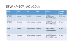
Management of IUGR Management of IUGR Fetal monitoring Fetal monitoring Monitoring the growth-restricted fetus involves serial fetal measurement -Abdominal circumference - Amniotic fluid index - Cardiotocography - Doppler ultrasound *Fetuses with absent end-diastolic flow are hypoxaemic, these changes may appear up to 5 weeks before demise *Reversed end-diastolic flow is suggestive of preterminal compromise; the fetus may die within 1-2 days if not delivered. Amniotic Fluid Index Amniotic Fluid Index -Fetal urine production is significantly lower in the SGA fetus than in the AGA fetus. - Decreased renal perfusion results in oligohydramnios Biophysical Profile Biophysical Profile -Breathing - Tone - Movement - Amniotic fluid volume Cardiotocography * Requires about 40 mins observation of fetal breathing movements. (Takes time not for every fetus only suspect IUGR) -A persistently abnormal biophysical score is associated with absence of end-diastolic flow
Management of IUGR Management of IUGR *The objective of clinical management is to expedite delivery before the occurrence of fetal compromise, but after fetal lung maturation has been achieved. *The timing of delivery should be based on the results of the antenatal testing, fetal growth pattern, dopplers, and gestational age. *For women with antiphospholipid antibodies associated with the delivery of a prior IUGR infant, low-dose aspirin (81 mg/day) in early pregnancy may reduce the likelihood of recurrent IUGR. *For cases in which ultrasonic findings strongly suggest IUGR, with or without abnormal fetal surveillance, delivery is indicated at gestational ages of 34 weeks or later, or at any reasonable gestational age if pulmonary maturity is documented. *At birth, the infant should be carefully examined to rule out the possibility of congenital anomalies and infections. The monitoring of blood glucose levels is important because the fetuses do not have adequate hepatic glycogen stores, and hypoglycemia is a common finding.
prevention prevention Risks All women should be encouraged to stop smoking since it is the commonest risk factor - Even passive smoking is harmful husbands should be persuaded to stop. - Early aspirin treatment before 17 weeks (100-150mg) for patients with previous IUGR babies (possible role of placental thrombosis) LOW DOSE! Neonatal: Polycythemia ,Hyperbilirubinemia , Hypoglycemia , Hypothermia , Apneic episodes . Intrapartum: Increased risk of fetal heart rate abnormalities , Cesarean delivery , Low Apgar scores , Cord blood acidemia. Long term: Largely dependent on the etiology of the IUGR and the gestational age at delivery , Lifelong increased risk of cardiovascular disorders , insulin resistant, obesity.
Done by: Sarah alshehri Revised by: Razan AlDhahri


")