Intravenous Therapy: Scope, Indications & Anatomy
Delve into the world of intravenous therapy with insights on the scope of practice for infusion nurses, common indications for IV therapy, and the anatomy of skin and veins. Learn about the crucial skills and knowledge required for safe and effective IV administration.

Download Presentation

Please find below an Image/Link to download the presentation.
The content on the website is provided AS IS for your information and personal use only. It may not be sold, licensed, or shared on other websites without obtaining consent from the author. If you encounter any issues during the download, it is possible that the publisher has removed the file from their server.
You are allowed to download the files provided on this website for personal or commercial use, subject to the condition that they are used lawfully. All files are the property of their respective owners.
The content on the website is provided AS IS for your information and personal use only. It may not be sold, licensed, or shared on other websites without obtaining consent from the author.
E N D
Presentation Transcript
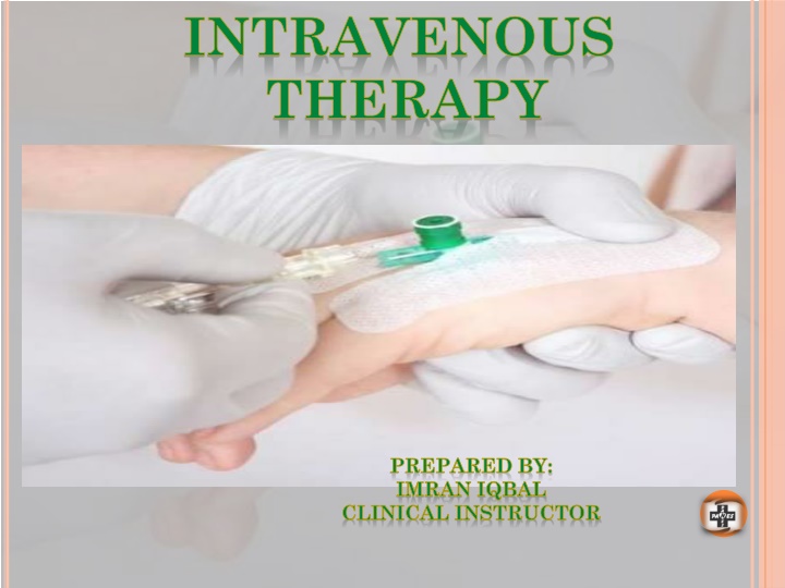
INTRAVENOUS THERAPY PREPARED BY: IMRAN IQBAL CLINICAL INSTRUCTOR
SCOPE OF PRACTICE . The infusion nurse is accountable for practicing within the defined scope of practice for the RN and is committed to provide safe and quality infusion nursing care.
SCOPE OF PRACTICE . The infusion nurse s practice is based upon the following: Knowledge of anatomy and physiology. Specific knowledge and understanding of the vascular system and its relationship with other body systems. Knowledge of infusion treatment modalities. Participation in the establishment of the patient s ongoing plan of care.
SCOPE OF PRACTICE . Skills necessary for the administration of IV therapy. (Latest Technologies) The infusion nurse should perform nursing process. The process should include :- Assessment. Problem identification/ Nursing diagnosis. Outcome identification. Planning and communication.
SCOPE OF PRACTICE . Implementation and evaluation. The infusion nurse should collect: Data. Prioritize patient problems and needs. Develop and implement care plan. Evaluate patient outcomes.
INDICATIONS OF INTRAVENOUS THERAPY
INDICATIONS OF IV THERAPY The purpose for IV insertion must be assessed prior to its insertion. (Equipments, Site, Patient s Teaching)
INDICATIONS OF IV THERAPY Some of the Indications are: To administer medications. To replace fluids and electrolytes. To increase caloric intake when GI tract is not a viable choice. Blood and blood products Administration. To administer TPN ( Total Parental Nutrition) To Administer diagnostic reagents (e.g. Contrast dyes) To monitor hemodynamic functions.
ANATOMY OF SKIN AND VEIN
ANATOMY OF SKIN AND VEIN The first barrier to successful peripheral IV insertion is skin. Barrier between outside environment and internal organs. The risk for the infection increases whenever skin is broken. An infusion access device perforates the skin, interrupts the integrity of the barrier and increases the risk of infection. sterile Technique is very important for IV insertion, care and maintenance.
ANATOMY OF SKIN AND VEIN The Skin consist of two main layers: 1-EPIDERMIS. The epidermis is the first defence against infection. It is tough protective layer that contains melanin ( which protects against the rays of the sun and give the skin its colour)
ANATOMY OF SKIN AND VEIN 2- DERMIS. Much thicker and directly below the epidermis. Dermis layers consists of blood vessels, hair follicles , sweat glands, sebaceous glands, collagen and nerves. Human skin is only about 0.07 inches (2mm) thick. About 2 square meter, which weighs about 2.7kg
ANATOMY OF SKIN AND VEIN 3-VEIN. An understanding of the anatomy of vein and venous system will assist the nurse in selecting the appropriate site for IV insertion. A vein consist of three layers. Tunica Adventitia. (The outer most layer) Tunica Media. ( The Middle layer) Tunica Intima ( The inner most layer)
SELECTION OF VEIN FOR IV INSERTION
SELECTION OF SITE FOR IV INSERTION Proper selection of vein is key to successful IV therapy. Points to be remembered while selecting the site: Suitable location. Patient preference. Amount and type of medication to be infused. Expected duration of IV therapy. Accessibility of vein.
SELECTION OF SITE FOR IV INSERTION Size and condition of the vein. Patient s age, size, general condition, and preference. Presence of disease or surgery (e.g. Mastectomy) Presence of shunt and graft. Your experience and skill at venipuncture. Use a systematic approach ( begin distally and work proximally) Choose veins which are long, easily palpable and visible.
SELECTION OF SITE FOR IV INSERTION RENAL PATIENT: Avoid the extremity with the shunt and graft. POST MASTECTOMY: Avoid the operative arm, Avoid arms with burns, Scars, Paralysis etc. Avoid cannulation if there is indication of skin infection, pain or trauma. Avoid veins which are hard with cord-like feeling.
SELECTION OF SITE FOR IV INSERTION ELDERLY PATIENT: Skin on the hands and dorsal metacarpal veins are often fragile, avoid such veins. LOWER EXTREMITIES: Are more susceptible to complications ( Doctor s Ordered required) Diabetic Patients: Should never be cannulated on lower extremities.
SELECTION OF SITE FOR IV INSERTION JOINTS FLEXION AREAS: Avoid it can increase risk of mechanical damage. Avoid veins close to arteries or arterial lines e.g. antecubital fossa. Avoid previously cannulated veins- intima may be damaged. Avoid small, visible but impalpable veins.
EQUIPMENTS NEEDED FOR IV CANNULATION Followings are the equipments needed for IV cannulation. IV cannula. Tourniquet. Antiseptic Solution (2% Chlorhexidine or (70% alcohol or 10% Povidone iodine) Sterile 2-by-2 Gauze. Saline Flush. Transparent Dressing / Tape. IV fluids Bag with tubing. Volume control device. Sharp Container.
EQUIPMENTS NEEDED FOR IV CANNULATION
EQUIPMENTS NEEDED FOR IV CANNULATION
EQUIPMENTS NEEDED FOR IV CANNULATION
EQUIPMENTS NEEDED FOR IV CANNULATION .
EQUIPMENTS NEEDED FOR IV CANNULATION
EQUIPMENTS NEEDED FOR IV CANNULATION
EQUIPMENTS NEEDED FOR IV CANNULATION
PROCEDURE OF PERIPHERAL CANNULATION Assemble all the equipments. Explain the procedure to the Patent. Prepare the patient for procedure. Observe universal standard precautions. Hand washing. Apply clean gloves. Assess and choose a vein. Choose appropriate needle. If needed clip the hair at site but don t shave.
PROCEDURE OF PERIPHERAL CANNULATION Disinfect the skin at the insertion site. (circular motion ) Allow the site to dry. Do not palpate the vein after disinfecting. Injection lidocaine if needed. Apply tourniquet, tight enough to distend the vein. Holding the skin taunt. Insert the needle or catheter through the subcutaneous tissue at the angle of 10-20 degree.
PROCEDURE OF PERIPHERAL CANNULATION As the needle enters the vein assess for blood return indicating successful venipuncture. If you don t see blood return, release the tourniquet and discontinue the attempt. Do not exceed more than two attempts. If the blood return present, attach the syringe with normal saline and release the tourniquet. Gently flush the tubing and check for infiltration. Secure the catheter with transparent occlusive dressing over it.
PROCEDURE OF PERIPHERAL CANNULATION Don t cover the actual insertion site. Label the IV site with, Type Gauge Date and time of insertion Initials of the inserting person. For infants and paediatric patient use a splint or arm board to avoid dislodgement of IV Cannula. Assess for circulation and prevention of pressure.
MAJOR STEPS IN MAINTAINING PERIPHERAL IV Double check the physician order. Check the appropriateness of the solution orders. Assess the rate to be infused. Any IV additives should be labelled clearly along with: Patient s name Room Number Additives name and dosage Volume per hour
MAJOR STEPS IN MAINTAINING PERIPHERAL IV Drip rate Date and time the solution was hung. Nurses signature Assemble all the equipments needed Assess the IV solution to be infused for: Clarity Cracks Leaks Date of expiry
MAJOR STEPS IN MAINTAINING PERIPHERAL IV Assess the IV tubing for kinks and tight connections. Calculate the infusion rate. Three question must be answered prior to the rate. How much solution did the physician order? Total time of infusion? What is drop factor of IV tubing ? Assess the IV site for any signs of local complications. Documents the procedure. A peripheral IV has to be discontinued if a physician orders. If any Complication has occurred at the site. IV should be changed every 72 hours to avoid phlebitis.
DISCONTINUATION OF PERIPHERAL IV PROCEDURE: Check physician s order. Wash Hands. Turn off the IV fluids. Ensure aseptic techniques. Put on Non-sterile gloves. Stabilize the needle with one hand, while removing the dressing. Gently withdraw the needle at the same angle it was inserted. Immediately apply pressure with dry sterile gauze till bleeding stops. Avoid using alcohol swabs, it can delay clotting time. Assess the site and apply a band aid or dressing .
DISCONTINUATION OF PERIPHERAL IV PROCEDURE: Document the procedure and appearance of site. If the site appears infected, a swab should be taken and sent with tip of the cannula for culture and sensitivity.
DOCUMENTATION Documentation should must contain complete information regarding: Infusion therapy. Insertion of peripheral cannula. Complications. Adverse reactions. Documentation must be done according to the organizations guideline.
DOCUMENTATION INSERTION OF IV CANNULA Evidence of informed consent. Reason for insertion of IV cannula. Date and time. Details of site preparation. Numbers of attempts made in insertion of cannula. Insertion site including name of the vein. Name of the person placing the cannula. Problems encountered during insertion. Appearance of site after insertion, (Bruising/bleeding) Type of Dressing.
DOCUMENTATION INSERTION OF IV CANNULA Local anaesthesia used. Flush solution used/Amount. Functionality of cannula immediately after insertion, e.g. presence of blood return/ability to flush device easily. Type, size and gauge of catheter device. Patient s response and tolerance to procedure. Overall condition of the patient.
DOCUMENTATION OF ONGOING CARE / MAINTENANCE Details of catheter care. Site care and condition. Appearance (Standardized local assessment scales) Assessment of insertion site for: Redness Edema Rashes Discoloration Drainage Intactness
DOCUMENTATION OF ONGOING CARE / MAINTENANCE Flush solution, ( Volume, Frequency, Difficulties) Specify safety and infection control precautions taken. Catheter replacement.
DOCUMENTATION OF INFUSION THERAPY Clear and accurate detailed record of IV medication. Type of therapy administered: Drug Rate Route Time Method of administration.
DOCUMENTATION OF INFUSION THERAPY Pertinent diagnosis Assessment and monitoring of vital signs Patient s tolerance Response to therapy Symptoms and appropriate laboratory tests/ results Record any adverse drug reactions Any adverse events Complications of therapy / Peripheral cannula Record result of any monitoring
DOCUMENTATION OF INFUSION THERAPY Record site assessment Record the administration of IV drugs/fluids
DOCUMENTATION OF COMPLICATIONS RELATED IV THERAPY Document any complications. Side effects of infusion therapy. Date time and situation when complication was noted. Complications noted with peripheral cannula. Strategies used to manage the complications. Evaluation of effectiveness. Documentation of extravasations incidents.