Introduction to Digital Radiography and PACS
Digital imaging in radiology involves processes that create electronic images for viewing and manipulation on computers. Digital radiography uses indirect and direct capture methods to convert x-rays into images viewable on computer screens. PACS systems store, retrieve, distribute, and present medical images in DICOM format, facilitating seamless integration of various imaging instruments. DICOM standardizes information handling in medical imaging, enabling interoperability among different hardware from various manufacturers.

Download Presentation

Please find below an Image/Link to download the presentation.
The content on the website is provided AS IS for your information and personal use only. It may not be sold, licensed, or shared on other websites without obtaining consent from the author.If you encounter any issues during the download, it is possible that the publisher has removed the file from their server.
You are allowed to download the files provided on this website for personal or commercial use, subject to the condition that they are used lawfully. All files are the property of their respective owners.
The content on the website is provided AS IS for your information and personal use only. It may not be sold, licensed, or shared on other websites without obtaining consent from the author.
E N D
Presentation Transcript
Introduction To Digital Radiography And PACS By : Alanoud Al Saleh
Digital Imaging Digital imaging is defined as any image acquisition process that produces an electronic image that can be viewed and manipulated on a computer. In radiology, images can be sent via computer networks to a variety of locations.
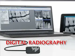
Digital Radiography Two types of digital radiography Indirect capture DR : Machine absorbs x-rays and converts them to light. Uses of charge-coupled device CCD or thin-film transistor (TFT) converts light to electric signals. Computer processes electric signals. Images are viewed on computer monitor.
Digital Radiography Direct capture DR Photoconductor absorbs x-rays. Thin-film transistor TFT collects signal. Electrical signal is sent to computer for processing. Image is viewed on computer screen.
Picture archiving and communication system In medical imaging, picture archiving and communication systems (PACS) are computers or networks dedicated to the storage, retrieval, distribution and presentation of images. The medical images are stored in an DICOM format (Digital Imaging and Communications in Medicine).
DICOM (Digital Imaging and Communications in Medicine) (DICOM) is a standard for handling, storing, printing, and transmitting information in medical imaging. It includes a file format definition and a network communications protocol.
DICOM files can be exchanged between two entities that are capable of receiving image and patient data in DICOM format. DICOM enables the integration of scanners, servers, workstations, printers, and network hardware from multiple manufacturers into (PACS).
Types of images in PACS Most PACSs handle images from various medical imaging instruments, including ultrasound, magnetic resonance, PET, computed tomography, endoscopy, mammograms, etc.
PACS uses Reading stations Physician review stations Web access Technologist quality control stations Administrative stations Archive systems Multiple interfaces to other hospital and radiology systems 9