Introduction to Enterobacteriaceae and E. coli in Microbiology
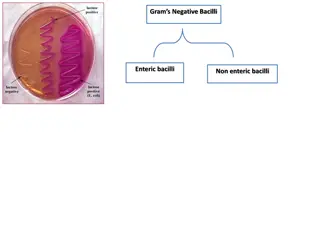
This content explores the characteristics, morphology, and growth patterns of Enterobacteriaceae, a large group of Gram-negative rods commonly found in various environments. It delves into the identification of typical organisms, culture characteristics, and biochemical tests used for differentiation. Differential media such as EMB and MacConkey agar are highlighted for distinguishing lactose fermenting and non-fermenting enteric bacteria.

Download Presentation

Please find below an Image/Link to download the presentation.
The content on the website is provided AS IS for your information and personal use only. It may not be sold, licensed, or shared on other websites without obtaining consent from the author.If you encounter any issues during the download, it is possible that the publisher has removed the file from their server.
You are allowed to download the files provided on this website for personal or commercial use, subject to the condition that they are used lawfully. All files are the property of their respective owners.
The content on the website is provided AS IS for your information and personal use only. It may not be sold, licensed, or shared on other websites without obtaining consent from the author.
E N D
Presentation Transcript
University of Mustanisiriya College of Medicine Department of microbiology Introduction To Enterobacteriaceae and E. coli Dr Ali Abdulwahid
The Enterobacteriaceae Overview Large group of Gram-negative rods Found worldwide in soil, water, and vegetation. Some are part of the normal intestinal flora of animals and humans Others are pathogenic for humans eg. salmonellae and shigellae, Involve many genera : Escherichia, Shigella, Salmonella, Enterobacter, Klebsiella, Serratia, Proteus, and many others.
Common characteristics Members of this family share these common characteristics: i. Gram-negative bacilli. ii. Aerobes or facultative anaerobes iii. All species ferment rather than oxidize glucose with the production of acid or acid and gas. iv. Either motile with peritrichous flagella or non-motile. v. Catalase positive vi. Oxidase-negative vii. Grow well on MacConkey agar
Morphology and Identification I. Typical Organisms 1. Short gram-negative rods 2. Some species possess a capsule such as Klebsiella species (large and regular capsules), whereas other species lack the capsules. II. Culture E coli and most of the other species form circular, convex, smooth colonies with distinct edges. Enterobacter colonies are similar but more mucoid. Klebsiella colonies are large and very mucoid The salmonellae and shigellae produce colonies similar to E. coli but do not ferment lactose. Some strains of E. coli produce heamolysis on blood agar.
Growth charecteristics The organisms of this family display different patterns of carbohydrate fermentation and amino acid decarboxylation and other enzymes, which are used in biochemical differentiation IMVIC test : The four biochemical tests widely employed in the classification of enterobacteria are , which involve : the indole (I) , methyl red (MR), Voges- Proskauer (VP) and citrate utilization tests (C). triple sugar iron (TSI) agar, https://microbeonline.com/imvic-tests-principle-procedure- and-results/
Culture on differential media such as ( eosin- methylene blue [EMB], MacConkey agar) are used to differentiate between lactose fermenting (LF) and none lactose fermenting enteric bacteria These media containing carbohydrates (such as lactose ) and special dyes as pH indicators (such as methylene blue) MacConkey agar with LF and non-LF colonies By Medimicro - Own work, Public Domain, https://commons.wikimedia.org/w/index.php?curid=4511724
Antigenic Structure Antigenic variability can be used for serogrouping the enteric bacteria Three major antigens involved : 1. O antigens (somatic antigens): polysaccharide chains in the lipopolysaccharide complex (LPS) of the outer membrane 2. H antigens (flagella antigens) consisting of protein located on flagella 3. K antigens (capsular): Linear polymers of the outer membrane consists of a repeated units of carbohydrate (sometimes proteins as well) The bacteria can be detected in agglutination assays with specific antibodies against these antigens the antigenic formula of an E. coli can be O55:K5:H21.
Main Virulence factors I. Surface antigens : 1. Somatic antigen (O Antigen) oExerting endotoxic activity oalso protects the bacteria from phagocytosis and the bactericidal effects of human complement system . 2. K Antigen oProvide protection against phagocytosis and antibacterial activity of human normal serum 3. Fimbriae oAct as adhesion factor that promotes adhesion to human tissue such as in colon, bladder, and others . oProtect the bacteria from being washed away in the UT.
II.Toxins 1. Endotoxins: The toxicity of the lipid A portion of LPS of outer membrane responsible for many of the systemic manifestations of infection. 2.Exotoxins : such as haemolysins, enterotoxins and cytotoxines Usually lead to cause watery diarrhea III.Antibiotic resistance factors (their encoding genes often are carried on plasmids and can be transferred between related species .
Medically important species 1- Opportunistic organisms: E. coli, Klebsiella, Enterobacter, Proteus species, Morganella, Providencia, Citrobacter, and Serratia They are members of the normal intestinal microbiota. Non-invasive They cause diseases only when they transfer into other body sites or tissues outside their normal intestinal sites or other less common intestinal sites When normal host defences are inadequate, localized clinically important infections can result, and the bacteria may reach the bloodstream and cause sepsis.
2- Pathogenic organisms Salmonella, Shigella, and Yersinia species They are invasive to human and always cause diseases
Escherichia coli (E. coli ) Morphology is a gram-negative, straight rods arranged singly or in pairs. It is nonsporing motile by peritrichate flagella, though some strains may be non-motile. http://www.bacteriainphotos.com/Escherichia%20coli %20light%20microscopy.html
Cultural Characteristics Cultural Characteristics Colonies are large, thick, grayish white, moist, smooth, opaque or partially translucent disks. Many pathogenic isolates have polysaccharide capsules. Some strains may occur in the mucoid form. On blood agar, many strains, especially those isolated from pathogenic conditions, are hemolytic. On MacConkey s medium, colonies are red or pink due to lactose fermentation. In broth, growth occurs as general turbidity and a heavy deposit, which disperses completely on shaking. Source of picture : http://www.bacteriainphotos.com/cultivation%20media/MC%2 0larger/escherichia%20on%20macconkey4.jpg
Biochemical Reactions E. coli ferments glucose, lactose, mannitol, maltose and many other sugars with the production of acid and gas. Typical strains do not ferment sucrose. IMVIC test : Indole and MR positive, and VP and citrate negative (+ + - - ) It is also negative for urease test, H2S production, and gelatin liquefication.
Antigenic Structure Serotyping of E. coli is based on three antigens (as in other entric bacteria: 1. the flagellar antigen H 2. somatic antigen O 3. the capsular antigen K
Main virulence factors 1- surface antigens : Including : H, O and K antigens 2- toxins : including a) endotoxins b) exotoxins: two types: 1. Hemolysins 2. Enterotoxins (usually cause diarrhea) : There are three distinct types enterotoxins produced by E. coli i. Heat labile toxin (LT) ii. Heat stable toxin (ST) iii. Verotoxin (VT) also known as Shiga-like toxin (SLT).
Heat labile toxin (LT) Heat Stable Toxin (ST) A small, monomeric toxin binds to and activates guanylate cyclase in the enterocytes increase in the level of cyclic guanosine monophosphate cGMP subsequent hypersecretion of fluid Like cholera toxin Multimric toxin (1 A subunit + 5 B subunits) Activates adenyl cyclase in the enterocyte increase the level of cyclic adenosine 5 monophosphate (cAMP), Increased outflow of water and electrolytes into the gut lumen, with consequent diarrhea. Shiga-like toxin (SLT). Comprise one A and five B subunits. Penetrate the cells and subsequently disrupts protein synthesis. Destruction of the intestinal villus results in decreased absorption with a relative increase in fluid secretion.
Diseases 1. Urinary tract infection E. coli is the most common cause of UTI 2. Sepsis The bacteria may reach the bloodstream and cause sepsis in: Immune-compromised people Newborns Sepsis can also occur as a secondary infection to UTI. 3. Meningitis E. coli along with group B streptococci are the leading causes of meningitis in infants.
4. E.coliassociated diarrheal diseases E. coli strainsthat cause diarrhea are classified according to their virulence properties and their mechanisms in causing diseases into five groups: Such groups involved : Enteropathogenic E. coli (EPEC) Enterotoxigenic E. coli (ETEC ) Enteroinvasive E. coli (EIEC) Enteroaggregative E. coli (EAEC) Shiga toxin-producing E. coli (STEC)
i. i. Enteropathogenic Enteropathogenic E. coli E. coli (EPEC) (EPEC) One of the leader causes of diarrhea in infants in developing countries. The infection is associated with severe watery diarrhea; vomiting; and fever. The infection is usually self-limited but can be prolonged or chronic. The duration of diyarrhea can be shortened and the chronic infection can be treated by antibiotics
ii. ii. Enterotoxigenic Enterotoxigenic E. coli E. coli ( (ETEC ETEC) ) The common cause of traveler s diarrhea and a very important cause of diarrhea in infants in developing countries The bacteria initially adhere to the mucosal surface of the epithelial cells of the small intestine. It produces a heat-stable enterotoxin (ST) or a heat-labile (LT) cholera like enterotoxin, or both The infection is waterborne or foodborne It is usually of brief duration, involving a rapid onset of loose stools along with variable symptoms , including nausea, vomiting and abdominal cramps. Antimicrobial prophylaxis can be effective Antibiotic treatment effectively shortens the duration of diyarrhea.
iii. iii. Enteroinvasive Enteroinvasive E. coli E. coli (EIEC) (EIEC) The organisms invade epithelial cells of the large intestine, penetrate them and multiply intracellularly, leading to blood and mucus in the stool. Spreading from one cell to neighbouring cells leads to tissue destruction and consequent inflammation , leading to symptoms of bacillary dysentery Infections are usually food-borne but there is also evidence of cross-infection between persons (faecal-oral). The pathogenesis and clinical picture of EIEC infections are ranging from mild diarrhea to frank dysentery, and occurs, in children as well as adults.
iv. iv. Enteroaggregative Enteroaggregative E. coli E. coli (EAEC) (EAEC) They are able to adhere to particular laboratory-cultured cells in an aggregative or stacked brick pattern causes acute and chronic diarrhea (>14 days in duration) in developing countries. cause of foodborne infection in developed countries associated with traveller s diarrhea and persistent diarrhea in patients with HIV. produce a low molecular weight heat stable enterotoxin called EAST ( enteroaggregative heat stable enterotoxin-1) Some isolates express hemolysins. Symptoms : watery diarrhea, vomiting, occasional abdominal pain, dehydration >14 days
v. v. Shiga toxin Shiga toxin- -producing producing E. coli E. coli (STEC) (STEC) The strain is so called Shiga toxin-producing E coli (STEC) due to the cytotoxic toxins they produce It is associated with haemorrhagic colitis (a severe form of diarrhea) and with haemolytic uremic syndrome Infection either sporadic or occur as out breaks of food poisoning (in the community, nursing homes of elderly and day care centres) Direct and in direct Contamination with human or animal faeces is the main source of infection Infection can be prevented by thoroughly cooking meat and avoiding unpasteurized products
Treatment and prevention Treatment Antibiotic therapy must take into consideration the resistance pattern of the pathogen. Aminopenicillins, ureidopenicillins, cephalosporins, quinolones, or cotrimoxazole are useful agents. oral replacement of fluid and electrolyte losses are essential in severe diarrhea Prevention Eating thoroughly cooked foods and drink disinfected water is of important to prevent infection for travellers chemoprophylaxis with anti-infective agents can be used in preventing traveller s diarrhea
References 1. Reidle, S., Morse, S. A., Meitzner, T., and Miller, S. 2019. Jawetz, Melnick & Adelberg s Medical Microbiology , Twenty-Eight Edition. The McGraw-Hill education, Inc. USA 2. Kumar, S. 2012. Textbook of microbiology. Jaypee Brother Medical Publishers (P) Ltd. New Delhi, India. 3. Websites : 1. Microbiology in picture (https://www.microbiologyinpictures.com) 2. Bacteria in photos (http://www.bacteriainphotos.com) 3. Microbe online (https://microbeonline.com)
Thank You For Your Attention


")


")