Introduction to Urine Analysis in the Urinary System
The urinary system consists of kidneys and urinary tracts, each kidney containing nephrons. Urination is a voluntary process, and disturbances include painful urination, frequency, and urinary retention. Gross examination of urine involves physical examination parameters like urine volume and color, which can indicate various health conditions.

Download Presentation

Please find below an Image/Link to download the presentation.
The content on the website is provided AS IS for your information and personal use only. It may not be sold, licensed, or shared on other websites without obtaining consent from the author. If you encounter any issues during the download, it is possible that the publisher has removed the file from their server.
You are allowed to download the files provided on this website for personal or commercial use, subject to the condition that they are used lawfully. All files are the property of their respective owners.
The content on the website is provided AS IS for your information and personal use only. It may not be sold, licensed, or shared on other websites without obtaining consent from the author.
E N D
Presentation Transcript
Introduction to urine analysis Lec. 1
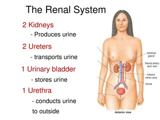
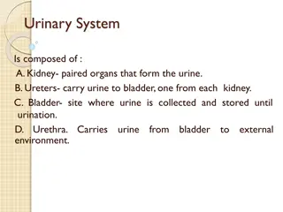
The urinary system Urinary system Urinary system consists of pair of kidneys and urinary tracts which includes (two ureters, urinary bladder and urethra). Each kidney contains 1.3 million urinary units called nephrons. Each nephron consists of glomerulus and urinary ducts (Bowman's capsule, proximal convoluted tubule, Henley loop, distal convoluted tubule and collecting duct).
Urination Urination: is a voluntary process depends on person's choice for suitable time and place to empty the urinary bladder from storage urine, without pain. Disturbances in urination include: 1- Painful urination, frequency, Urgency. Because of microbial UTI. 2- Impairment of urine flow, hesitancy, drubbing urine, incomplete emptying. Because of urinary bladder obstruction. 3- Urinary retention, a sign of benign prostate enlargement (hypertrophy), (enuresis) due to dysfunction of bladder muscles or sphincter muscles. urinary incontinence
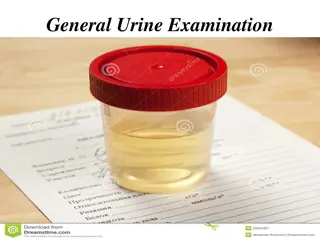
Gross examination of urine: Physical examination
Physical examination 1-Urine volume: This is dependent environmental condition, diet and activity of the human. Value above or below the normal value (1.5 L/Day) can be considered as pathological disorder but it should be combined with clinical and laboratory examination. - above normal (polyuria) urine volume (< 2.5-3L/Day) due to large quantities intake of liquids, diuretics, alcohol, in sufficient of urinary ducts in re-absorption of water and urine concentrated as in diabetes mellitus or diabetes insipidus. - under normal (Oligourea) urine volume (< 400 ml/Day) - Anuria, urine volume (< 50 ml/Day), due to: hot weather, sweating, low water intake, or due to disease in kidney or urinary ducts. normally up on fluid intake,
2- Color: Can be observed in a test tube or in a urinometer tube. Yellow to amber (Normal); the color comes primarily from the presence of urobilin. Urobilin is a final waste product resulting from the breakdown of heme from hemoglobin during the destruction of aging blood cells. Colorless to pale yellow; dilute urine with low specific gravity and polyuria. Dark yellow or yellow brown; concentrated urine with a high specific gravity and small quantity. Yellow brown or greenish yellow; yellow green foam when urine is shaken.Urobilinoids chromagon derived from heme green biliverdin yellow-brown-billirubin-and urobilin. Cloudy; hematuria (clearer after centrifugation). Translucent; hemoglobinuria. Brown to brownish black; hemoglobin up on standing bile large amounts. Green; bile biliverdin Red to pink; phenothiazine (beet root) Blue; medication contain methylene blue or food with blue dyes.
3-Transparency (clarity): Clear freshly voided urine is clear. Cloudy not necessarily pathological as many samples may become cloudy. Epithelial cells present in large numbers. Blood red to brown color and smoky. Leukocytes may appearance if large number. Bacteria produce a uniform turbidity if in large number; the turbidity doesn t settle out and cannot be removed by filtration. produce milky, ropy
4-Specific gravity (SG): Determined by refractometer and indicator paper stripes. Normal value in Man : 1.010 1.030 (Average normal = 1.025). depending on SG the urine will be diluted (SG 1.002) or concentrated (SG 1.065) depend on the solvents in urine. SG used to determine the kidney efficiency keeping water balance in urine.
5-Odour: Normal odor aromatic or acetone Abnormal odor aromatic odor of ketone bodies (sweet fruit odor) as in starvation and diabetes. In UTI the odor of urine is bad smell. 6-Foam: Shake the sample and observe: If the amount of foam produced is in excess and slow to disappear proteinuria If the color of the foam great yellow or bile pigments If the color of the foam red to brown hemoglobinuria brown
Microscopic Examination of Urine Sample It is of great clinical importance and should never been omitted. Important structure to be include: Casts: Cylindrical bodies performed in distal collecting tubules from RBCs or WBCs or fatty compounds or waxes. Diagnosis of cast type aid in diagnosis of the disease. Types of casts: (Hyaline casts, density granulated casts, finely granulated casts, red cell casts (hematouria), and (inflammation), wax epithelial casts. white casts, cell casts, casts and fat
Erythrocytes (RBC) and leukocytes (pus cells): present in urine in case of UTI, diseases, and inflammation. Epithelial cells: presence normally due to sloughing the lining layer of urinary tubules, bladder and urethra or because of some renal diseases. Budding yeasts: Candida albicans, found in diabetes patients urine because of the low PH and the presence of sugar necessary for the growth. Protozoa: like Trichomonas vaginalis that infect the vagina in women and urethra in men and cause trichomoniasis. Mucus: mucus is a slimy secretion that originates from the mucous membranes and glands Crystals: a salt compounds organized in geometrical shapes and looks like crystals, important in stones formation (cysteine crystals and oxalate crystals). Amorphous urate white or pink cloud in acid urine Amorphous phosphate white cloud in alkaline urine
Chemical Examination of Urine: -PH of urine The normal hydrogen ion (pH) concentration, in the urine (5-8) depends on the type of diet. Vegetable diet, citrus fruits (also bacterial infections) produce alkaline urine, while high protein diet (also blood acidosis where PH 7.35, some microbial infections, ketones elevation due to diabetes or aspirin intake) produce acidic urine. PH measured by paper strip or pH meter. -Proteins of urine: a little quantity, of protein are found normally in urine (150 mg/day)any access in protein called proteinuria which is an indication for many diseases like kidney diseases , fever and pregnancy.
Types of protein in urine: 1-Albumine: is the first protein appearing in urine due to its low molecular weight and size (albuminuria), this protein appears in Diabetes and hypertenation. 2- Immunoglobulins: appear in urine due to inflammations and microbial infections 3-Hemoglobine: found in urine due to blood hemolysis. Test: (Robert s test) Principle: Precipitation of protein by strong acid A positive test is indicated by a white ring at the zone of concentration, which should be read against a dark back ground and reported as: Negative - No ring at the zone of concentration Note: In many clinical laboratories, Robert s test is routine method as it is simple, quick and easy to read even when only a small amount of protein presents.
Glucose in urine: No glucose is present in the urine normally which passes glomerular filter, because it is completely absorbed in the tubules. It present when the blood glucose level elevated to(180mg/ml) which is called renal threshold, when blood glucose elevated the glucose present in urine as in diabetes. Glucose Test Method: Qualitative method (Benedicts test) Principle: Reducing sugars present in the urine react with the copper sulphate to reduce the cupric ions to cuprous oxide giving a Color change from blue (negative) to green, yellow and red depending on the amount of reducing substances present


:")
:")


 and leukocytes (pus cells): present")
 and leukocytes (pus cells): present")