Iron Deficiency Anemia (IDA) Overview

Iron Deficiency Anemia (IDA) is a prevalent condition with significant impacts on health and quality of life. Learn about its background, definition, epidemiology, and etiology, along with the diagnostic algorithm and key laboratory findings for identification. Explore the different classifications and understand the process of red blood cell production and maturation.

Download Presentation

Please find below an Image/Link to download the presentation.
The content on the website is provided AS IS for your information and personal use only. It may not be sold, licensed, or shared on other websites without obtaining consent from the author.If you encounter any issues during the download, it is possible that the publisher has removed the file from their server.
You are allowed to download the files provided on this website for personal or commercial use, subject to the condition that they are used lawfully. All files are the property of their respective owners.
The content on the website is provided AS IS for your information and personal use only. It may not be sold, licensed, or shared on other websites without obtaining consent from the author.
E N D
Presentation Transcript
Iron Deficiency Anemia (IDA) Ass.prof.Abeer Anwer
Objectives Explain background, definition, epidemiology, and etiology of iron deficiency anemia (IDA) Outline diagnostic algorithm of IDA Identify key laboratory findings to diagnose IDA
Background Anemia is a group of disease characterized by a decrease in either hemoglobin (Hb) or circulating red blood cells (RBCs) o Results in reduced oxygen-carrying capacity of the blood According to World Health Organization (WHO) o 1.6 billion people (1/4 of world s population ) are anemic Not an innocent bystander; affects both length and quality of life (QOL) IDA occurs across all populations and is associated with o Diminished QOL o Physical and cognitive performance, and o Unfavorable clinical outcomes
Morphological Classification Anemia Macrocytic Normocytic Microcytic Genetic Anomaly Megaloblastic Non-megaloblastic IDA 1. 2. Hepatic disease Drug-induced anemia Hypothyroidism Reticulocytosis 1. Vitamin B12 deficiency Folic acid deficiency 1. 2. Sickle cell Thalassemia 1. 2. 3. 4. Recent blood loss Hemolysis Bone marrow failure Anemia of chronic disease 2. 3. 4.
Definition According to WHO o Anemia is defined as Hb <130 g/L in men or <120 g/L in female IDA is the result of long-term negative iron balances o Progressive loss of iron stores in the form of hemosiderin and ferritin IDA is defined as o Anemia with biochemical evidence of iron deficiency based on following laboratory findings Serum ferritin, total iron binding capacity (TIBC), transferrin saturation, or transferrin receptor
Epidemiology IDA is the most common nutritional deficiency in developing and developed countries IDA is considered to be the leading cause of anemia worldwide, accounting for as many as 50% of cases Prevalence of IDA greatly varies according to age, gender, physiological, pathological, environmental, and socioeconomic conditions prevalence of IDA o Young children 1.2% o Women of childbearing age 4.5%
Process of RBC Production & Maturation RBC production Iron + Hb
Iron Metabolism: Figure 16-8: Iron metabolism
Recent in iron metabolism the TMPRSS6 gene encoding Matriptase-2. Matriptase-2 (MT-2).. is a transmembrane serine protease that cleaves Hemojuvelin, a major regulator of hepcidin expression and plays an essential role in down-regulating hepcidin, the key regulator of iron homeostasis. When blood iron levels are low, this signaling pathway reduces hepcidin production, allowing more iron from the diet to be absorbed through the intestines and transported out of storage sites (particularly in the liver and spleen) into the bloodstream
Erythropoietin is the specific erythroid growth factor. It is produced in the kidneys by peritubular interstitial cells in response to hypoxia. It acts on erythropoietin receptors on the surface of developing erythroid cells. The expression of this receptor declines rapidly with maturation of the cells. Hemoglobin consists of four hem & four globin chains. The hem consists of a protoporphyrin ring with ferrous iron in the center. The globin chains consist of 2 & 2 non chains. The main hemoglobin in post natal life is HbA ( 2 2). Also present in adults HbA2 ( 2 2) up to 3.5% & HbF ( 2 2) up to 1%.
Iron transport: two atoms of ferric iron can attach to a single molecule of transferrin which is synthesized by the liver. Normally transferrin is one third saturated & it gains most of the iron from the macrophages of the RE system which degrade hemoglobin after death of old red cells (not from the GIT). The developing erythroblasts are rich in transferrin receptors so they get most of transferrin iron. Storage iron: ferritin is water soluble iron storage protein. It is synthesized by the liver. One molecule can bind 4000 iron atoms. Hemosiderin is water insoluble & is formed by partial degradation of ferritin. It is visible in the macrophages by Prussian blue stain (iron stain)
Iron Balance Normal iron content of the body o 3-4 g (Hb, myoglobin, and cytochromes) Iron is best absorb as ferrous (Fe2+) form in the duodenum, and to a smaller extent in jejunum Daily recommended allowance o Adult males/postmenopausal females: 8 mg o Menstruating female: 18 mg Iron sources o Heme iron (2-3X more absorbable): meat, fish, and poultry o Non-heme iron: vegetables, fruits, dried beans, nuts, grain products, and dietary supplements Gastric acid/ascorbic acid increases non-heme iron absorption whereas phytates (in bran), tannins/polyphenols (in tea), and calcium (in dairy product) form insoluble complexes
Pathophysiology Iron stores are reduced without reducing serum iron levels and can be assessed with serum ferritin measurement Initial Stage Iron stores can be depleted without causing anemia Once iron stores are depleted, there still is adequate iron from daily RBC turnover for Hb synthesis Second Stage Iron deficiency occurs; Hb falls just above the lower limit normal Third Stage Considered as IDA and occurs because of Hb falls to less than normal values
Etiology IDA results from prolonged negative iron balance Mainly due to following factors: Inadequate iron intake Decreased iron absorption Increased iron demand or hematopoiesis Increased iron loss 1. 2. 3. 4.
Common Cause Age & Sex Females in the reproductive period of life Menstruation Pregnancy Pathological blood loss Deficient diet Adult males and postmenopausal females Pathological blood loss Infants and children Deficient diet Diminished iron stores at birth Etiology
ETIOLOGY The most important cause world-wide is infestation with parasitic worms (hookworms- suck 0.03- 0.2 ml of blood per worm /day ),whipworms, roundworms Dietary insufficiency Malabsorption
Prognosis IDA adversely effects o Cognitive performance, behavior, and physical growth of infants, preschool, and school-aged children o The immune status and morbidity from infections of all age groups o The use of energy sources by muscle and thus the physical capacity and work performance of adolescents and adults of all age groups o Increase perinatal risks for mothers and neonates and overall infant mortality during pregnancy
Preliminary Findings Chief Complaints Fatigue, lassitude, palpitation, and generalized weakness History Chronic blood loss, deficient diet Clinical Features 1. Palor skin, nailbed, conjunctiva 2. Koilonychia (brittle, spoon shaped nails) 3. Atrophic glossitis (atrophy of tongue papilla; making the tongue smooth and shiny) 4. Pica (compulsive eating of nonfood items) or pagophagia (compulsive eating of ice)
Signs & Symptoms Symptoms Signs Decreased exercise tolerance Tachycardia Pale appearance (most prominent in conjunctiva) Fatigue Dizziness Decreased mental acuity Increased intensity of some cardiac valvular murmurs Irritability Weakness Palpitations Vertigo Shortness of breath Chest pain
Laboratory Evaluation Complete blood count (CBC), erythrocyte sedimentation rate (ESR), and peripheral blood film (PBF) Serum Iron profile Bone marrow study (if needed) Investigations to determine other causes of IDA (e.g. fecal occult blood test, colonoscopy, urine examination)
Hematological Parameters in IDA Hematologic Indices IDA Hb Low Hematocrit (Hct) Low Mean corpuscular volume (MCV) Low Mean corpuscular hemoglobin (MCH) Low Mean corpuscular hemoglobin concentration (MCHC) Low Red cell distribution width (RDW) High (early)
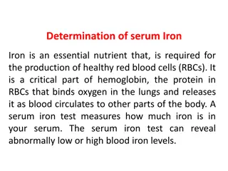
Iron Profile Lab Exams Comments In IDA 1. 2. 3. 4. It is the concentration bound to transferrin Approximately one-third transferrin bound to iron Levels are decreased by infection and inflammation Best interpreted in conjunction with TIBC Serum Fe (50-100 mcg/dL) Low 1. Ferritin (storage iron) is proportional to total iron stores Best indicator of iron deficiency or overload Infection or inflammation can increase the concentration, independent of iron status Serum ferritin (>10-20 mcg/L) Low 2. 3. 1. Indirect measurement of the iron-binding capacity of serum transferrin (protein) 2. Levels don t fluctuate over hours or days unlike serum iron Total iron binding capacity (TIBC) (250-410 mcg/dL) High 1. 2. Ratio of serum iron level to TIBC in percentage Reflects the extent to which iron-binding sites are occupied on transferrin and indicates the availability of iron for erythropoiesis Less sensitive and specific for IDA than ferritin % Saturation of transferrin (>20%) Low 3.
DiagnosisLABORATORY INVESTIGATIONS Bone marrow aspiration, with the marrow stained for iron - Bone marrow is hypercellular, with erythroid hyperplasia Leukocytes and megakaryocytes are normal No stainable iron in marrow reticulum cells
Summary IDA is the most common form of anemia and is usually the result of prolonged negative iron balance in the body Four main factors contributing to IDA include o Inadequate iron intake o Decreased iron absorption o Increased iron demand or hematopoiesis o Increased iron loss Clinical diagnosis of IDA should include complete patient history and physical exams, followed by laboratory investigations Abnormal laboratory investigations generally include low MCV, serum iron, and ferritin; and high TIBC
Summary Treatment of IDA usually consists of dietary supplementation and administration of oral iron preparations General recommendation for oral iron replacement is 200 mg elemental iron/day, divided into 2-3 doses to maximize tolerability Parenteral therapy is usually not indicated unless patient is intolerant to oral therapy, having malabsorption, or in the case of CKD Anaphylactic reaction should be considered for all parenteral formulation along with strictly monitoring adverse drug reaction
Summary Decision to manage anemia with blood transfusion is based on the evaluation of the risk and benefit and is only considered when Hb is <70 to 80 g/L Complete therapeutic response requires iron supplementation for up to 2-6 months, however, symptoms may improve within few days after oral therapy
Self Assessment Questions Q1: Which of the following is one of the common cause of IDA in young male? Deficient diet Menstruation Pathological blood loss None of the above A. B. C. D.
Self Assessment Questions Q2: Microcytic hypochromic anemia can be due to the following factor(s): Folic Acid Vitamin B12 C. Iron deficiency Hemolysis A. B. D.
Self Assessment Questions Q3: Which of the following statement is false regarding iron in our body? It is best absorb in ferrous (Fe2+) form in the duodenum Heme iron is found in meat, fish, and poultry Gastric acid/ascorbic acid increases non-heme iron absorption Non-heme iron is 2-3X more absorbable than heme iron A. B. C. D.
Self Assessment Questions Q4: Identify the following laboratory investigations for diagnosing IDA as high/low: Hb Low MCV Low Serum iron Low TIBC High Serum ferritin Transferrin saturation Low Low
Self Assessment Questions Q5: For oral iron products, the following statements are true except: Ferrous sulfate tablet contains 65 mg elemental iron Administration of oral iron should be 1 hour before meals or on empty stomach preferably Can cause GI intolerance and discoloration of stools Percent elemental iron of all oral preparations is roughly the same A. B. C. D.