ISBMR Osteoporosis Diagnosis & Treatment Position Statement
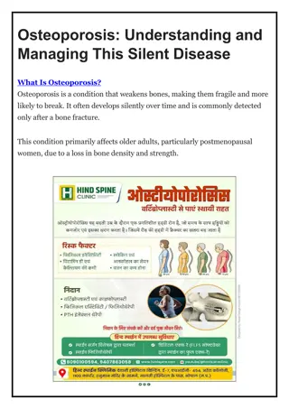
Position paper by ISBMR highlights the need for guidelines on osteoporosis management in India. The paper addresses the lack of accurate data, increased prevalence due to aging population, urbanization, and low awareness. It emphasizes the challenges in accessing diagnostic tools and treatment, underscoring the importance of developing strategies to combat osteoporosis in the Indian population.

Download Presentation

Please find below an Image/Link to download the presentation.
The content on the website is provided AS IS for your information and personal use only. It may not be sold, licensed, or shared on other websites without obtaining consent from the author.If you encounter any issues during the download, it is possible that the publisher has removed the file from their server.
You are allowed to download the files provided on this website for personal or commercial use, subject to the condition that they are used lawfully. All files are the property of their respective owners.
The content on the website is provided AS IS for your information and personal use only. It may not be sold, licensed, or shared on other websites without obtaining consent from the author.
E N D
Presentation Transcript
The Indian Society for Bone and Mineral Research (ISBMR) position statement for the diagnosis and treatment of osteoporosis in adults Subhash C Kukreja, MD University of Illinois Chicago, IL, USA
Position Paper vs. Guidelines Position Paper vs. Guidelines A Position Paper is a detailed policy report, drafted by members of a society, that explains or advocates a certain course of action. It s commonly derived from research support studies. Practice Guidelines are systematically developed statements based on the best evidence and the most current data. These guidelines are ideal for helping both practitioners and patients make healthcare decisions in specific circumstances and help standardize medical care and improve the quality of care. We don t yet have enough data from India to come up Guidelines for Osteoporosis management
Epidemiology of Osteoporosis Population in India is aging and surviving longer and therefore prevalence of osteoporosis will increase in coming years. However, there are no accurate data available on the incidence or prevalence of this disease in India. Most data on the prevalence of osteoporosis among women in India come from studies conducted in small groups spread across the country, and estimates from 2015 have suggested that 20% of the 230 million Indian women over age 50 have osteoporosis [4, 5]. Prevalence of osteoporosis ranging from 8 to 62% in Indian women of different age groups has been reported in several studies [6 14]. The prevalence of osteoporosis in males older than 50 years is also variable, ranging from 8.5 to 24.6% [9, 15, 16]. A 2001 study in expatriate Indians in Singapore showed that the incidence of hip fracture in the Indian population was 361 for women and 128 for men per 1,00,000 population
Epidemiology Cont. Urbanization appears to be associated with an increased prevalence of osteoporosis due to lifestyle habits such as sedentary lifestyle, increased indoor living, and lower sun exposure [4]. The awareness of osteoporosis is low in India, with surveys indicating that only 10 15% of Indians are aware of the disease [17]. According to the International Osteoporosis Federation, the availability of dual-energy X- ray absorptiometry instruments (DXA), a key tool for diagnosing osteoporosis, is about 0.26 per million in India, far below the recommended number of 10.6 per million [18]. Moreover, most of the DXA instruments are located in urban areas, and even many large cities in India do not have DXA facilities. Furthermore, the fact remains that the cost of DXA and osteoporosis treatments are largely not covered by insurance. India fares poorly compared to the more developed Asian countries like Japan and Korea, where availability of DXA is much higher (20.8 and 24.5 per million, respectively) [19] Low awareness of disease and poor availability of DXA are among factors associated with not having a good handle on osteoporosis in India
Risk Factors for Osteoporosis Risk Factors for Osteoporosis (No different than other populations) populations) (No different than other
Race/Genetics Race/Genetics Generalizing all Indians as a single race assumes uniformity of genetics across the population. It is becoming clear that Race (as either self-assigned by patient or a physician) is not an easily defined concept and may be a poor surrogate for genetics Although the average age at menarche in Indian girls is ~ 12.5 years, the average age at menopause is 46.2 years which is earlier than that seen in non-Indian women [22], and this is a significant risk factor for the development of osteoporosis in Indian women [13, 23]. Genetic factors, race, and ethnicity also have a major influence on peak bone mass attainment. Asian Indian women have been shown to have 5 15% lower bone mineral density (BMD) than non-Asian women [24 26]. Also, polymorphisms in the gene for vitamin D receptors in different races have been suggested to contribute to the ethnic differences in BMD [4, 27, 28].
Data base for defining Normal BMD Data base for defining Normal BMD Most of the data on fracture/BMD relationship has been derived using young Caucasian women as the reference population. The 2019 International Society for Clinical Densitometry (ISCD) Official Position recommends the use of a uniform Caucasian (non-race adjusted) female normative database for women of all ethnic groups. It also states that manufacturers should continue to use NHANES III data as the reference standard for femoral neck and total hip T-scores [49]. Although normative data on BMD in healthy Indian adults exist [9, 50, 51], at present, there is insufficient data to assess the fracture risk using the Indian BMD reference database. Hence, for all practical purposes and in line with the 2019 ISCD Official Position, the Caucasian female database derived from the NHANES III is used as India s reference population for calculating T-scores [52]. Caucasian female data base is the most validated in terms of its relationship to defining fractures. Both mean values and SD need to be considered for defining normal ranges. Potential adjustments can be considered especially in small- frame individuals (e.g. using -3.0 as a cut off rather than -2.5 for T-Score)
Nutritional Factors Nutritional Factors Calcium: Indian diets are predominantly vegetarian, and the contribution of dairy products to the overall calcium intake is minimal in the lower socioeconomic classes. Furthermore, the unequal distribution of milk and milk products, with boys and men being served larger portions, is another factor that worsens the situation [5]. Indian diets have a higher ratio of phytates to calcium, especially among rural Indians [30]. Phytates may hinder calcium absorption from the already calcium-deficient diets. A survey conducted in 2011 2012 in India reported a dietary calcium intake of only 429 mg/day [31]. Vitamin D: Some of the reasons for vitamin D deficiency among Indians may be lower sun exposure due to indoor lifestyle, traditional clothing leading to less skin exposure to sunlight (saris, salwar-kameez,etc.), inadequate dietary intake, poor vitamin D fortification of foods, and darkly pigmented skin and atmospheric pollution [18, 35]. Vitamin D deficiency results in ineffective calcium absorption from the gut, which in turn affects the mineralization of bones. The findings of the Delhi Vertebral Osteoporosis Study (DeVOS) from India suggest that the odds of having osteoporotic fractures in subjects consuming calcium and vitamin D supplements are lower [36]. Poor Nutritional status: Leading to low body mass and sarcopenia Urbanization: Leading to lower exercise and poorer sun exposure Diabetes Mellitus: There is increasing prevalence of type 1 and type 2 Diabetes Mellitus, increasingly recognized as a risk factor for osteoporosis.
Diagnosis of Osteoporosis Diagnosis of Osteoporosis Any adult with a fragility fracture should be suspected of having underlying osteoporosis (primary vs. secondary). In addition, historical height loss of more than 4 cm in postmenopausal women raises the possibility of asymptomatic vertebral fractures [45]. Dual-energy X-ray absorptiometry, or DXA, is the most commonly used technique for measuring BMD. Although true density measurement is 3- dimensional, DXA is a two dimensional measurement and thus calculates areal bone density [46]. We recommend against the use of ultrasound (QUS) for screening or initial decision-making regarding treatment of osteoporosis. Diagnosis of osteoporosis is made by history of fragility fracture and/or low BMD by DXA; DXA being the gold standard tool despite its limitations
Indian Indian- -Specific FRAX Specific FRAX Derivation of Indian-specific FRAX is based on the fracture risk in individuals living in Singapore and, therefore, may not be fully applicable in the native Indian population. Using the NOF treatment cut-off guidelines of 3% risk for hip fracture and 20% risk for MOF, Indian-specific FRAX may underestimate the fracture risk [68]. Based on studies of Indian patients with hip fractures, these thresholds may be lower, and studies are underway to define these lower treatment thresholds [58, 69]. Trabecular bone score (TBS), a tool that assesses bone s microarchitecture, has emerged as a valuable modality complementary to BMD [70, 71]. A pan-India reference for TBS is underway; however, therapeutic guidelines and thresholds cannot currently be based on TBS [73]. FRAX Tool as currently published for the Indian population, is not reliable and its use will likely lead to under-treatment. Attempts are underway to revise the Tool. However, until accurate data are available for hip fracture incidence in the Indian population (?regional), the tool will likely remain unreliable. The 3% (Hip) and 20% (MOF) guidelines are based on an economic model in USA and not necessarily applicable to Indian population
Osteoporosis Risk Assessment Tools Several osteoporosis screening tools have been validated in both women and men in the Indian population. These tools are based on simple clinical risk factors and could be easily used in community settings [74]. In a large cohort of rural postmenopausal women, the SCORE (Simple Calculated Osteoporosis Risk Estimation) screening tool was useful, with good sensitivity and good area under the curve for predicting femoral neck osteoporosis on BMD measurement. It uses simple clinical risk factors like age, weight, previous fracture, estrogen therapy, rheumatoid arthritis, and ethnicity. [75]. Similarly, the risk assessment tools OSTA (osteoporosis self-assessment tool for Asians)and MORES (male osteoporosis risk estimation score) have been validated for use in the Indian population [76]. These tools are rapid, easy to perform, inexpensive, and easily usable in the rural Indian setting, but the impact of initiating therapy based on thresholds derived from these tools is not well studied. Best use of Risk Assessment tools is to identify individuals who would benefit most from DXA measurement
Osteoporosis Screening Recommendations Osteoporosis Screening Recommendations Since data indicates that osteoporotic fractures occur at an earlier age in Indians than in the West, we recommend screening at an earlier age [5, 55]. Women aged 60 and older and men aged 65 and older, regardless of clinical risk factors Postmenopausal women younger than 60 years and men aged 60 64 years when there are concerns for osteoporosis based on their clinical risk factor profile Women in the menopausal transition if there is a specific risk factor associated with increased fracture risk, such as low body weight, prior low-trauma fracture, or high-risk medication Individuals who have had a fragility fracture before the age of 50 years Individuals with a condition (e.g., rheumatoid arthritis, diabetes mellitus, malabsorption syndrome) or who are taking medication (e.g., glucocorticoids in a daily dose 5 mg prednisone or equivalent for 3 months) associated with low bone mass or bone loss Any individual being considered for pharmacologic therapy for osteoporosis Other than the screening to start at an earlier age, these recommendations are similar to those published for other populations
Recommendations Biochemical Investigation Recommendations Biochemical Investigation These recommendations are similar to those published for other populations
Bone Turn Over Markers Bone Turn Over Markers Bone turnover markers (CTxand P1NP) are dynamic parameters that reflect short-term, acute changes in bone remodeling status that are not measured by BMD and hence, are complementary to BMD measurement. However, BTMs have no role in the diagnosis of osteoporosis. Although BTMs are not routinely used to diagnose osteoporosis, they are increasingly used in the follow-up of patients who are on anti-osteoporotic treatments. Hence, wherever available, patients contemplating anti-osteoporotic therapy can get a baseline BTM level estimated prior to initiation of therapy for subsequent comparison during follow-up [57]. CTx and/or PINP can be used to evaluate patient adherence and drug responses to anti-resorptive agents, with measurements suggested at baseline, 3, 6, and 12 months after starting treatment. Similarly, PINP can be used to evaluate patient adherence and drug responses to anabolic agents, with measurements at baseline, 1 to 3 months, 6 months, and 12 months after starting anabolic treatment. Bone Turnover Markers may be useful in monitoring compliance and response to therapy but not for diagnosis of osteoporosis
Indications for Osteoporosis therapy Indications for Osteoporosis therapy Indian-specific formula for FRAX noted above is still a work in progress and needs validation. It is recognized that DM ( Type 1 or Type 2) is an important risk factor for osteoporosis
Life Style and Nutrition in OP Management These recommendations are similar to those published for other populations
Recommendations for initial first Recommendations for initial first- -line therapy for individuals with prevalent vertebral fractures for individuals with prevalent vertebral fractures line therapy Teriparatide is an effective anabolic agent to initiate therapy in these cases, which to be continued for 24 months and followed by anti- resorptives. Intravenous zoledronic acid or denosumab are also effective options. Since the protocol for discontinuing denosumab is still not firmly established, zoledronic acid is usually preferred as initial therapy for 3 5 years. Oral bisphosphonates can be used if the patient wants to avoid injectable therapies.
Recommendations for initial first Recommendations for initial first- -line therapy for individuals with prevalent hip fracture for individuals with prevalent hip fracture line therapy Intravenous zoledronic acid is the agent of choice in this group it is recommended that hospitalized/postsurgical patients with hip fracture be given a dose of intravenous zoledronic acid before being discharged from the hospital. Denosumab is also an apt and effective choice but is often used after zoledronic acid(concerns for rebound vertebral fractures after stopping drug).
Recommendations for initial first Recommendations for initial first- -line therapy for high for high- -risk individuals without prevalent fractures risk individuals without prevalent fractures line therapy Bisphosphonates are generally agents of choice for those at high risk for fracture. While either weekly oral (alendronate, risedronate) or annual intravenous agents are effective, concerns about compliance and ease of once a year administration has made zoledronic acid the preferred drug for most patients. Both options should be discussed with the patient (weekly oral vs. annual intravenous) and treatment chosen accordingly. Denosumab can be used as a first choice too if the patient reacts to or wants to avoid bisphosphonates. The risk of rebound fractures is increased, if subsequent doses of denosumab are not administered in time Teriparatide can be considered for some with very low BMD (T score < 3.5) and high risk of vertebral fracture..
Recommendations for the use of sequential Recommendations for the use of sequential therapies in the management of osteoporosis [79] therapies in the management of osteoporosis [79] Treatment with teriparatide should always be followed by anti- resorptive agents to prevent bone density decline denosumab can be used in this setting. In patients unresponsive to anti-resorptive therapy alone, treatment can be followed by a combination of teriparatide and anti- resporptives [81]. Treatment with denosumab, if it has to be discontinued, should be followed by bisphosphonate, either zoledronate [82] or alendronate [83] in patients with adequate renal function. Delay in denosumab therapy or lack of an alternate therapy 6 months after last denosumab dose is associated with a rebound increase in fractures.
Recommendations Recommendations- - Other Agents Other Agents Intranasal calcitonin can be used for temporary bone pain relief. However, calcitonin s effectiveness in prevention of osteoporotic fractures is very limited and should therefore be prescribed only in women who cannot tolerate bisphosphonates, denosumab, teriparatide, or raloxifene. Estrogens: Although effective in increasing bone mass and prevent fractures, HRT is not recommended for managing osteoporosis due to high risk of side effects such cardiac events and breast cancer (although breast cancer risk is not increased with estrogens alone). Hormone Replacment Therapy can be used when there is an additional indication to use estrogens such as uncontrollable menopausal symptoms. Testosterone therapy may be added in androgen-deficient men (testosterone level less than 200 ng/dL on more than one determination) if accompanied by signs or symptoms of androgen deficiency These recommendations are similar to those published for other populations
Recommendations for the management of osteoporosis in chronic kidney disease patients and those on hemodialysis Management of patients with osteoporosis and chronic kidney disease (CKD) is difficult as bisphosphonates are contraindicated in stage 4 and 5 kidney disease (eGFR below 30 to 35 mL/min). Denosumab is not cleared by the kidney and therefore can be used in these patients. However, the risk of hypocalcemia is high with this agent, especially in patients in stage 5 disease. Optimal calcium intake and vitamin D status should be assured before starting denosumab. A major concern with antiresorptive therapy in patients with CKD is adynamic bone disease and selected patients should undergo undecalcified iliac bone biopsy if facilities are available, to guide correct decision-making for the management of osteoporosis
Drug Holiday Drug Holiday The concept of a drug holiday has been proposed to potentially reduce the incidence of the rare adverse events associated with long-term anti-resorptive therapy [99]. However, the recommendation for drug holidays is still a matter of debate [100], especially since there is a dearth of data from India. However, the Endocrine Society guidelines 2020 do recommend a drug holiday in selected groups of patients [59]. In patients on bisphosphonate therapy, fracture risk needs to be evaluated after 3 5 years (3 years for intravenous, 5 years for oral therapy). however, fracture risk needs to be evaluated regularly at 2 4 year intervals, with therapy being reinstituted if the patient falls into the high-risk category. In patients on denosumab therapy, fracture risk needs to be evaluated in 5 10 years. A drug holiday can be considered in low-moderate risk patients following a course of bisphosphonate with fracture risk being revaluated every 1 3 year. There is no consensus on using BTMs to assess the need for drug holiday [95].
Follow Up of Patients Follow Up of Patients There exists no consensus regarding the frequency of follow- up for patients on anti-osteoporotic therapy. The first follow-up can be planned after 3 months following initiation of therapy. Thereafter, patients can be followed up at 3 6 monthly intervals for 2 3 subsequent contacts followed by annual visits [21]. At each visit, a brief history with an emphasis on assessing new incident fractures, new-onset/worsening of kyphosis/ scoliosis, new-onset or worsening of back pain, and perceptible height loss should be elicited. A history of falls isa predictor of future falls and hence should be specifically queried. Patients should also be asked about the possible side effects of anti-osteoporotic therapy, notably, thigh and jaw pain. A short physical examination focusing on the patient s height should be undertaken. Other characteristics to assess include spinal tenderness, kyphosis, decreased spacing between lower ribs and pelvis, and oral hygiene. Patients on anti-resorptive therapy with poor dentition may be referred to a dental physician for a detailed oral evaluation. Biochemical testing- serum 25 OHD, renal function, serum calcium; initially 3 to 6 months intervals and then yearly X-rays of spine- annually as appropriate DXA monitoring- every 1 to 3 years as appropriate Bone Turnover markers- may help to assess compliance/response and effective of the drug.
Summary Summary Diagnosis of osteoporosis can be made in a patient with minimal trauma fracture without the aid of any other diagnostic tools. In others, bone mineral density measured by dual-energy X-ray absorptiometry remains the modality of choice. Data indicates that osteoporotic fractures occur at an earlier age in Indians than in the West; hence, screening for osteoporosis should begin at an earlier age. FRAX can be used for fracture risk estimation; however, it may underestimate the risk of future fractures in our population and still needs validation. Maintaining adequate calcium intake and optimum serum 25-hydroxyvitamin D levels are essential, which, in most cases, would require regular calcium and vitamin D supplementation. Pharmacotherapy should be guided by the presence/absence of vertebral/hip fractures or the severity of risk based on clinical factors, although bisphosphonates remain the first choice in most cases. Regular follow-up is essential to ensure adherence and response to therapy.
")
")