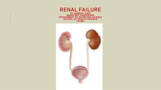
Kidney Injuries Overview
Learn about renal injuries, including causes, pathology, classification, and late complications. Blunt trauma is a common mechanism, leading to minor or major renal injuries. Vascular injuries are rare but serious. Detect potential late pathologies following renal trauma.
Download Presentation

Please find below an Image/Link to download the presentation.
The content on the website is provided AS IS for your information and personal use only. It may not be sold, licensed, or shared on other websites without obtaining consent from the author. If you encounter any issues during the download, it is possible that the publisher has removed the file from their server.
You are allowed to download the files provided on this website for personal or commercial use, subject to the condition that they are used lawfully. All files are the property of their respective owners.
The content on the website is provided AS IS for your information and personal use only. It may not be sold, licensed, or shared on other websites without obtaining consent from the author.
E N D
Presentation Transcript
INJURIES TO KIDNEY Renal injuries are the most common injuries of the urinary system. The kidney is well protected by ribs, vertebral bodies, lumbar muscles and the visera. Blows or falls on the loin and crushing road traffic accidentsare the usual causes of the injury to the kidney. Fractured ribs and transverse vertebral processes may penetrate the renal parenchyma.
Aetiology (i) Blunt trauma to the abdomen, flank or back is the most common mechanism of renal injuries. (ii) This accounts for 85% of all renal injuries. Such trauma may occur in motor accidents, fights, falls or in contact sports. (ii) Gun-shot and knife wounds may cause penetrating injuries to the kidney. Any such injury in the back or in the flank should be well examined to exclude renal injury. Associated abdominal visceral injuries are present in majority (80%) of such penetrating wounds. (iii) Sudden break in high speed vehicle may result in major vascular injury of the kidney.
PATHOLOGY AND CLASSIFICATION Blunt trauma usually causes laceration of the kidney in the transverse plane. Tears of the renal parenchyma usually follow the lines of the collecting tubules. The pathologic classification of renal injuries is as follows: (a) MINOR RENAL INJURY. Renal contusion or bruising of the parenchyma is the most common lesion. This minor renal trauma in fact constitutes majority (85%of cases) of renal injuries.
(b) MAJOR RENAL INJURY. This means deep corticomedullary lacerations which extend into pelvis to cause extravasation of urine in the perirenal space . This constitutes only 14% of cases or slightly more. The various types in this category are : (i) Complete fissure or tear of the renal parenchyma and pelvis to cause gross hematuria. (ii) Deep laceration of the kidney which causes large retroperitoneal and perinephric hematomas. (iii) Multiple lacerations causing complete destruction of the kidney. (iv) Laceration of the renal pelvis
(c) VASCULAR INJURY - Vascular injury of the renal pedicle is rare but may occur due to blunt trauma or sudden break in motor car accident. The vascular injury is rare and constitutes only 1% or less of all renal injuries. Various types of injury in this category are : (i) Stretch on the main renal artery without avulsion( injury by trauma) may cause renal artery thrombosis. (ii) There may be partial avulsion of the segmental branch of the renal artery (iii) Total avulsion of the renal artery or vein. The most important feature of vascular injuries is that it is difficult to diagnose and if this is not made quickly, it results in total destruction of the kidney.
LATE PATHOLOGIES. If the patient survives, late pathologies may develop from various types of 1. Hydronephrosis. Hematomas and urinary extravasation( urine leaks from the urinary tract and collects in other body cavities, such as retroperitonium) in the extra peritoneal tissue may result in perinephric hydrosis leading to obstruction of the uretero-pelvic junction. This leads to hydronephrosis. 2. Urinoma (collection of urine when urine doesn t flow out of the urethra as it should). Persistent urinary extravasation from deep lacerations were not repaired may result in large perinephric renal mass which may become infected to cause abscess formation. This large perinephric renal mass is called urinoma. which
3. Renal hypertension. Fibrosis around renal artery may constrict it to cause renal hypertension. Blood flow in non-viable tissue due to injury is compromised, which also results in renal hypertension. 4. Arteriovenous fistula. This only results after penetrating injury involving renal artery and vein.
Clinical Features The history should include detail description of the accident. In case of penetrating injuries, the type of weapon should be interrogated and assessed. In case of gun-shot wounds, the type of gun should be questioned as high velocity bullets cause much more extensive damage than low velocity ones. Microscopic or gross hematuria following trauma to the abdomen or loin indicates injury to the kidney. The degree of renal injury does not correspond to the degree of haematuria. Gross haematuria may occur in minor renal injury, whereas mild haematuria can occur in major trauma. Presence of haematuria should not be taken lightly and it demands full evaluation of injury to the kidney. About 1/3rd of cases of renal vascular injury are not associated with hematuria. These cases are usually due to sudden breaks in motor car accidents.
SYMPTOMS The most common complaints are pain and haematuria following trauma. 1. Pain. Pain may be localised to one flank or over the whole abdomen. Pain may be due to fractured ribs or pelvic fractures and due to injury to other abdominal viscera. 2. Hematuria. As mentioned above this is an important feature of kidney injury. The patient often complains of haematuria following accident Haematuria may occur just after the accident, or may appear some hours after the accident, or it may be as delayed as between 3rd day to 3rd week after the accident.
kidney injury has been suspected, the patient must be followed up carefully. Such delayed hematuria is usually due to dislodgement of a clot. When hematuria is profuse, the patient may complain of clot colic, which is almost similar to ureteric colic due to calculus. 3. General abdominal distension may be complained of after one or two days of injury This generalized abdominal distension is called metcrorism . This is caused by retroperitoneal hematoma involving the splanchnic nerves. 4. Patient may present with a swelling in the loin after injury. This is due to perinephric hematoma or extravasation of urine. Normal concavity of the loin disappears.
Physical signs In general examinations hypovolemic shock or signs of high blood loss may be noted. In local examinations there may be ecchymosis or bruise in the loin or upper part of the abdomen or in the back. Fracture of lower ribs may be evident. Abdominal examination may reveal tenderness in the loin there may be diffuse tenderness. A large palpable mass represent large retroperitoneal hematoma or extravasation of urine. Occasionally if the peritoneum is ruptured blood or urine may enter the peritoneal cavity causing distension of the abdomen. Bowel sounds may then be absent.
Investigations 1. Urine examination. Microscopic or gross haematuria is revealed in urine examination. 2. Straight X-ray of the abdomen will disclose fracture of lower ribs or vertebral body or transverse process fracture. 3. Excretory urography is performed as soon as the intravenous lines are established and resuscitation has begun. It is mandatory to demonstrate first and foremost an intact functional kidney on the other side. This urogram will clearly define the renal outlines, cortical borders and ureters. 4. Nephrotomography is indicated if excretory urogram does not provide with the necessary informations.
5. Ultrasonography and retrograde urography are of little use initially in the evaluation of renal injuries. 6. Computed tomography (CT scan) is also proving to be an affective mean for staging renal trauma. 7.Arteriography determines major arterial or branch injuries.
TREATMENT TREATMENT. A. Emergency measures. The objectives of this are to treat shock and haemorrhage. The measures consist of (i) The patient must be hospitalised and lie flat in bed. Movement is disallowed. (ii) A sedative is immediately given in the form of injection pethidine or morphine, which is also pain killer. (iii) An appropriate antibiotic is started at the same time to prevent infection of the haematoma. (iv) Intravenous infusion is started which should be followed by blood transfusion. Hourly pulse and blood pressure charts should be maintained.
(v) Samples of urine are sent for examination. Microscopic or macroscopic presence of blood is also noted. (vi) Once the central venous pressure (CVP) has been normal, excretory urography (I. V.P) should be done urgently to know that the other kidney is normally functioning. B Local management. 1. BLUNT INJURIES. (a) As mentioned earlier MINOR RENAL INJURIES constitute 85% of cases. These usually do not require any operation. Bleeding stops spontaneously with emergency measures. The patient should be in bed rest for about 1 week till macroscopic haematuria has been absent. Further activities are also reduced for 1 month. (b) In case of MAJOR RENAL INJURIES there is persistent retroperitoneal bleeding, urinary
INJURIES TO THE URETER INJURIES TO THE URETER Ureteral injury is rare. It may occur in stabbing and gun-shot injuries, but in majority of cases ureter is injured by the surgeons (iatrogenic). Surgically ureter may be injured while operating for cancers in the cervix and uterus, for endometriosis and for inflammatory and malignant diseases of the sigmoid colon. The ureter is also rarely injured in those surgeries where speed is of paramount importance.
Ureter may be injured in operations e.g. emergency caesarean section or for ruptured ectopic pregnancy. Extensive lymph node dissection may also injure ureter. Endoscopic manipulation of a ureteral calculus with a stone basket may result in ureteral perforation. Passage of a ureteral catheter beyond an area of obstruction may perforate ureter.
CLINICAL FEATURES If the ureter has been ligated inadvertently during operation, the postoperative course is usually marked by temperature of 101 - 102 F as well as flank or lower quadrant pain. The patient may also complain of nausea, vomiting and distension of abdomen due to paralytic ileus. Sometimes ureterovaginal or cutaneous urinary fistula develops, which usually appears within first 10 days after operation.
It must be remembered that bilateral ureteral injury or ligation is manifested by postoperative anuria. If there is stab or gun-shot wound in the loin, penetrating injury to the ureter should be suspected This usually takes place in the midportion of the ureter.
INVESTIGATIONS 1. Microscopic haematuria is noticed in 90% of cases. 2. Straight X-ray is not of much help except it may demonstrate a large area of increased density in the pelvis or in the retroperitoneal tissue which may arouse suspicion. 3. Excretory urography is more valuable as it may show a diffuse shadow below the kidney on the injured side. It may show hydronephrosis and delayed excretion. 4. Ultrasonography may outline urinary extravasation. It will also outline hydro ureter due to ligation of the ureter.
TREATMENT All cases of penetrating injury demands exploration. When the injury is recognized at the time of surgery. In this case an attempt is made to restore continuity. If the ureter has been partially clamped or included in a ligature, the clamp is immediately removed or the ligature is quickly cut. This is followed by cystoscopic catheterization of the ureter. It should be removed after 1 week.
Ureteral injury is recognised after surgery.As mentioned earlier these cases are often recognised by continuous complain of aching in the loin and continuous fever for the early postoperative period Sometimes urinary leaks develop per vaginum or through abdominal wall usually between 6th and 10th postoperative days. If the case is detected within 48 hours of surgery, re-exploration is indicated.


