Laboratory Specimen Dissection and Handling Guidelines
Discover the essential procedures for dissecting and handling specimens in a laboratory setting, including gross room techniques, tissue types, and detailed dissection plans for various organs like eyes, brain, lungs, muscles, and more. Explore the intricacies of fixing, cutting, and processing different types of tissues for accurate diagnosis and evaluation.

Download Presentation

Please find below an Image/Link to download the presentation.
The content on the website is provided AS IS for your information and personal use only. It may not be sold, licensed, or shared on other websites without obtaining consent from the author. If you encounter any issues during the download, it is possible that the publisher has removed the file from their server.
You are allowed to download the files provided on this website for personal or commercial use, subject to the condition that they are used lawfully. All files are the property of their respective owners.
The content on the website is provided AS IS for your information and personal use only. It may not be sold, licensed, or shared on other websites without obtaining consent from the author.
E N D
Presentation Transcript
3 3 stage stage th Ms.c. Fadhela Nafi Alrawi Ms.c. Fadhela Nafi Alrawi
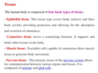
Type of tissues obtained in laboratory The human tissue comes from the surgery (Biopsy) and/or from the dissection room surgery two types of biopsy could be obtained: (Autopsy). From Incisional Biopsy: A small piece of lesions or tumor is removed and sent for diagnosis before final removal of the lesion or tumor. Excisional Biopsy: whole tumor or lesion is removed for examination.
Specimen dissection plans Small specimens need not to undergo any dissection and can be processed as they present ie whole ambedded. Eyes Eyes may be fixed in NBF usually for approximately 48 hours Brain this fixation takes 2-6 weeks. Fixation may also be enhanced by the use of microwave technology. Lungs Lung biopsies are typically fixed in NBF. Such fixed lungs can be cut within 2-6 hours Muscle biopsies The tissue for routine histological assessment is fixed in NBF and embedded so the fibers of the specimen are viewed in crosssection and longitudinally.
Specimen dissection plans Orient the specimen and cut parallel longitudinal slices 5 mm each For multinodularThyroid gland One section of each nodule (up to five nodules), including adjacent normal gland. More than one section for larger nodules Solitary Encapsulated Lesion For a solitary nodule measuring up to 5cm. take 1cm additional centimeter Thyroid gland-Thyroidectomy rim & encapsulated section for each
Specimen dissection plans Stomach _gastrectomy for tumer and ulcer : use formalin fixtive for 24 h
Specimen dissection plans Polypectomy: use formalin fixtive for 24 h Small bowel excision: use formalin or NBF fixtive for 24 h
Specimen dissection plans Appendix Divide specimen in three by cutting a cross-section 2 cm from tip Cut cross-sections of proximal fragment at 5 mm intervals kidney The sample is divided into three sections. It is taken from the affected area and from the lymph nodes, if any. Kidney nephrectomy for tumor
Specimen dissection plans Bladder_ cystectomy Two options are available for the dissection: Open with scissors in a Y shape through the anterior wall and fix formalin overnight in Fill with formalin, fix overnight and divide into anterior and posterior halves cutting the lateral bladder walls and then sectioning the prostate, beginning bladder neck and being careful to make the cut through the urethra. by first at the
Specimen dissection plans Prostate prostatectomy for tumor Serially section prostate at intervals from apex to base Lay the individual slices sequentially examine them carefully. Uterus - cervical biopsy gland: Radical the mm 2-3 and Do not cut the specimen unless the pieces are 4 mm It is essential that all of the tissue received preserved in formalin individual be
Specimen dissection plans Uterus hysterectomy Myomas: at least one section per myoma, up to three; sections from any grossly abnormal area (e.g. soft, fleshy, necrotic, cystic) Cervix: one section from anterior half and one from posterior half Cervical or endometrial polyps: to be submitted in entirety unless extremely large Hysterectomy For Malignant Tumours sample entire endometrium by making parallel sections, 2-3 mm apart,
Specimen dissection plans Fallopian tubes salpingectomy Submit three cross-sections of each tube, taken from the proximal, mid, portions, submitted same cassette and distal the in
Specimen dissection plans Breast: Lumpectomy / Excisional biopsy Section specimen: if specimen is s 3cm, cut 3-4 mm slices; if it is larger, bisect specimen transversely, place cut surface down, and take sagittal blocks through superior and inferior portions
GROSSING/SURGICAL CUT-UP Specimenreception: A separate room is required for specimen reception must be equipped with appropriate easily cleaned benching, adequate lighting, good ventilation, safetyequipment,disinfectants,absorptiongranulesandprotectiveclothing. Thekeypointofthisroomistoreceivesamplessafelyandsecurely. Any new specimen should have its identity confirmed and assigned a unique laboratoryspecimenidentifier,usuallyacomplexnumber. Mapping of the specimen identifier against the clinical request form is mandatory, along with checking of appropriate clinical details mentioned againstthespecimen. There must be facilities available that the pathologist can do the dissection eitherbystandingorsittingway.