Larynx Imaging Findings and Diagnostic Protocol
Explore various larynx imaging scan protocols, lesions, and conditions such as laryngocele, cancer types, and other abnormalities identified through MRI and CT scans. Understand the role of imaging in diagnosing and assessing laryngeal masses and related disorders.

Download Presentation

Please find below an Image/Link to download the presentation.
The content on the website is provided AS IS for your information and personal use only. It may not be sold, licensed, or shared on other websites without obtaining consent from the author. If you encounter any issues during the download, it is possible that the publisher has removed the file from their server.
You are allowed to download the files provided on this website for personal or commercial use, subject to the condition that they are used lawfully. All files are the property of their respective owners.
The content on the website is provided AS IS for your information and personal use only. It may not be sold, licensed, or shared on other websites without obtaining consent from the author.
E N D
Presentation Transcript
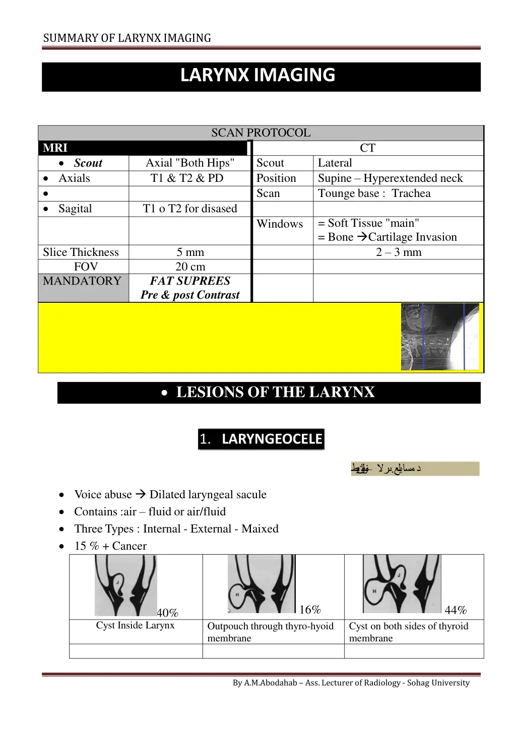
SUMMARY OF LARYNXIMAGING LARYNX IMAGING SCAN PROTOCOL MRI CT Scout Axials Sagital Axial "Both Hips" T1 & T2 & PD Scout Position Scan Lateral Supine Hyperextended neck Tounge base : Trachea T1 o T2 for disased = Soft Tissue "main" = Bone Cartilage Invasion 2 3 mm Windows Slice Thickness FOV MANDATORY 5 mm 20 cm FAT SUPREES Pre & post Contrast LESIONS OF THE LARYNX 1. LARYNGEOCELE . Voice abuse Dilated laryngeal sacule Contains :air fluid or air/fluid Three Types : Internal - External - Maixed 15 % + Cancer 16% 44% 40% Cyst Inside Larynx Outpouch through thyro-hyoid membrane Cyst on both sides of thyroid membrane By A.M.Abodahab Ass. Lecturer of Radiology - Sohag University
SUMMARY OF LARYNXIMAGING Air fluid level in External & Mixed 2. CANCER LARYNX Mostly Squamous cellcarcinoma Incidence 70 : 60% 25 : 35% 5 % Involve 3levels Lymphatic spread 30 % 1% 30 % Supra-glotic Glottic Sub Glottic Transglottic Role of Imaging: o CT & MRI Assess site & extension ofMass o In supraglottic lesions : Extension to pre epiglottic space & Para Glottic Space & Ant. Commisure By A.M.Abodahab Ass. Lecturer of Radiology - Sohag University
SUMMARY OF LARYNXIMAGING Lt Laryngeal mass invading Lt ary-epiglottic fold By A.M.Abodahab Ass. Lecturer of Radiology - Sohag University
SUMMARY OF LARYNXIMAGING 3. OTHERS HAMANGIOMA POLYP VoiceAbuse By A.M.Abodahab Ass. Lecturer of Radiology - Sohag University
SUMMARY OF LARYNXIMAGING By A.M.Abodahab Ass. Lecturer of Radiology - Sohag University
SUMMARY OF LARYNXIMAGING By A.M.Abodahab Ass. Lecturer of Radiology - Sohag University


