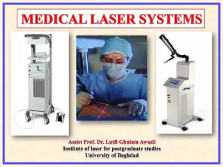
LASER in Ophthalmology: Principles and Applications
The physical principles and clinical applications of LASER technology in ophthalmology, covering photocoagulation, photodisruption, and more. Learn about LASER characteristics, parameters, types used in ophthalmic procedures, and indications for photocoagulation treatments like diabetic retinopathy. Enhance your understanding of LASER therapy in eye care."

Download Presentation

Please find below an Image/Link to download the presentation.
The content on the website is provided AS IS for your information and personal use only. It may not be sold, licensed, or shared on other websites without obtaining consent from the author.If you encounter any issues during the download, it is possible that the publisher has removed the file from their server.
You are allowed to download the files provided on this website for personal or commercial use, subject to the condition that they are used lawfully. All files are the property of their respective owners.
The content on the website is provided AS IS for your information and personal use only. It may not be sold, licensed, or shared on other websites without obtaining consent from the author.
E N D
Presentation Transcript
LASER in LASER in Ophthalmology Ophthalmology 2022 2022- -2023 2023 Dr. Zainab N. Hamoodi M.B.Ch.B, F.I.C.M.S(Ophth),ICO
Contents : Contents : Physical principles of LASER Clinical applications of LASER in ophthalmology : Photocoagulation Photodisruption Photoablation LASER classes Ocular LASER hazards LASER safety
LASER LASER L Light A Amplification by S Stimulated E Emission of R Radiation Characteristics of Laser : 1. Monochromatic = ONE color. 2. Coherent = all waves in the same phase. 3. Collimate = all waves are parallel. 4. High intensity.
Laser parameters : Laser parameters : 1. Power of the laser. 2. Pulse duration. 3. Spot size. 4. Number of shots. Types of laser used in ophthalmology : 1. Photocoagulation 2. Photodisruption 3. Photoablation
Photocoagulation The effect of laser depends on selective absorption of light energy by the ocular pigment Xanthophyll/Melanin/Hemoglobin Conversion to heat Elevation of tissue temperature to 60 C denaturate & coagulate cellular components. = Produce therapeutic burn to pre-selected area, with minimum damage to the surrounding tissues.
Types of laser used in photocoagulation : Argon laser Argon laser : medium = gas, it s of 2 types, Blue light (514 nm), green light (488 nm). Frequency doubled YAG laser Frequency doubled YAG laser : Medium = solid 532 nm Diode laser Diode laser : Medium = semiconductor red light (760 810 nm) Krypton laser Krypton laser : medium = gas red light Dye laser Dye laser : medium = fluid
Indications of photocoagulation : Treatment of diabetic retinopathy Panretinal phtocoagulation = PRP in proliferative diabetic retinopathy. Focal or grid laser photocoagulation in diabetic maculopathy. Treatment for retinal vein occlusion. Sealing of retinal breaks/holes. Treatment of open angle glaucoma argon laser trabeculoplasty (ALT) .
LASER photocoagulation in retinal vein occlusion
LASER photocoagulation for retinal beaks/holes
Photodisruption produce plasma tissue perforation / tissue cutting. ex. YAG laser (1046 nm), Femto laser(Nd:YAG 1053 nm) Indications of Indications of photodisruption photodisruption : : Peripheral laser iridotomy Posterior capsulotomy = thickening of the posterior capsule after cataract surgery. Cutting inflammatory membrane in front of intraocular lens. Cutting vitreous band. Cyclo-destructive = treatment of intractable glaucoma.
Post. Capsulotomy Peripheral iridotomy
Vitreolysis Cycloablation
Femtosecond laser Femtosecond laser Infrared laser, wavelength 1053 nm Nd:YAG laser, works by photodisruption Uses of femtosecond laser in ophthalmology: 1. Refractive surgery: Femtosecond Laser Assisted LASIK creating corneal flaps. Small Incision Lenticule Extraction (SMILE) 2. Femtosecond laser assisted cataract surgery creating corneal incision/ capsulorhexis.
Creating corneal flap in Femtosecond laser assisted LASIK
Femtosecond laser for creating corneal incision & capsulorhexis in cataract surgery
Photoablation Very short wavelength of UV light Interact & break chemical bonds of biological material. Tissue simply disappears without temperature rise. Excimer laser (139 nm) Indications of photoablation : Refractive surgery Eliminating refractive errors by changing the surface topography of the cornea. Removal of superficial corneal opacifications.
Laser Ocular Hazards These can result from either: Direct exposure to laser beam Specular reflection Reflection off diffused surface Harmful effects can be: Corneal burns Crystalline Lens damage Cataract Retina (Fovea) laser retinopathy
Laser Retinopathy The retina(especially fovea) is the most likely part of the eye prone for laser injury upon accidental exposure to high energy laser beam (without wearing protective goggles). sudden severe reduction in central vision, sometimes with scotoma. Laser retinopathy can be in the form of: Macular hole Macular pucker Macular hemorrhage Foveal burn
References Kanski clinical ophthalmology American Academy of Ophthalmology


")