Explore the pathology of lead toxicity in animals caused by ingestion of lead-based paints, improper handling of engine oil, and other sources. Learn about the absorption, distribution, and clinical effects of lead, including its impact on the nervous system, digestive system, and hematopoietic system. Discover the pathophysiology of lead toxicity and how it affects different organs and tissues in animals.
Please find below an Image/Link to download the presentation.
The content on the website is provided AS IS for your information and personal use only. It may not be sold, licensed, or shared on other websites without obtaining consent from the author. If you encounter any issues during the download, it is possible that the publisher has removed the file from their server.
You are allowed to download the files provided on this website for personal or commercial use, subject to the condition that they are used lawfully. All files are the property of their respective owners.
The content on the website is provided AS IS for your information and personal use only. It may not be sold, licensed, or shared on other websites without obtaining consent from the author.
E N D
Presentation Transcript
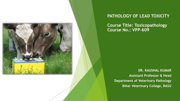
PATHOLOGY OF LEAD TOXICITY Course Title: Toxicopathology Course No.: VPP-609 DR. KAUSHAL KUMAR Assistant Professor & Head Department of Veterinary Pathology Bihar Veterinary College, BASU
Introduction Lead is a soft, malleable heavy metal. Lead poisoning is caused by increased levels of the heavy metal lead in the body tissues which can interfere with a variety of body processes and causes toxicity to many organs and tissue. Lead poisoning in animals and people is a major concern worldwide. It is most common in dogs and cattle. In other species such toxicity is limited by reduced accessibility, more selective eating habits and or lower susceptibility.
Sourses of poisoning Ingestion of lead-based paints Improper handling of engine-oil and Battery. Vegetation grown in lead smelter area. Feeding on crop sprayed with lead insecticides Deeping sheep in lead containing dip solutions (lead arsenate) as Sheep develop a kind of taste for lead.
Pathogenesis of Lead Toxicity Absorbed lead enters the blood and soft tissues and eventually redistributes to the bone. The degree of absorption and retention is influenced by dietary factors such as calcium or iron levels. Lead has a profound effect on sulfhydryl-containing enzymes, the thiol content of erythrocytes, antioxidant defenses, and tissues rich in mitochondria, which is reflected in the clinical syndrome. Lead interferes with essential metals as prosthetic groups of enzymes in the mitochondria, thus affects vital oxidative phosphorylation and ATP synthesis complex formation. This could explain the loss of integrity of nerve cell and functional disorder.
Pathophysiology of Lead Toxicity Lead salts (Carbonate /sulphate, tetraoxide) are sparingly soluble in water and hence 98% of ingested lead is eliminated in faeces. After absorption from GIT (1-2%), a large proportion is carried to RBC membrane. and rest either bind to serum albumin or remain free (unbound) which get distributed in soft tissues. Liver & kidney is the main organ where it gets deposited. It crosses the blood brain barrier and placental barrier as well. The bone is considered as SINK for lead as it may contain 90%-98% of the total body burden of lead.
CLINICAL FINDINGS Lead is irritating, immunosuppressive, gametotoxic, teratogenic, nephrotoxic, and toxic to the hematopoietic system. Nervous System Ataxia, salivation, tremor/ Spastic twitching of eyelids Champing of Jaw etc. Digestive System - Rumen stasis, colic, constipation diarrhoea, head pressing. Anemia is consistent finding in lead poisoning due to Destruction of erythrocytes (due to increased fragility) Depressant action on bone marrow (due to deposition of lead in bone). Inhibitor of heme synthesis. Chronic poisoning is seen in cattle and they show hyperaesthesia and other above mentioned signs.
LESION ACUTE LEAD POISONING : Macroscopic Lesion Oil or flakes of paint or battery may be evident in the GI tract. The caustic action of lead salts causes gastroenteritis. In the nervous system, edema, congestion of the cerebral cortex, and flattening of the cortical gyri on cerebrum are present. Microscopic Lesion Tubular necrosis in kidney, Centrilobular necrosis in liver and Osteopetrosis in affected animals
DIAGNOSIS Concentrations of lead in the blood at 0.35 ppm, liver at 10 ppm, or kidney cortex at 10 ppm are consistent with a diagnosis of lead poisoning in most species. Blood lead concentrations >0.05 0.10 ppm to be a notifiable disease in food-producing animals. Hematologic abnormalities, which may be indicative but not confirmatory of lead poisoning, include anemia, anisocytosis, poikilocytosis, polychromasia, basophilic stippling, metarubricytosis, and hypochromia. In dogs, rabies, distemper, and hepatitis may appear similar to lead poisoning. Canine blood smear, Basophilic Stippling in lead poisoning
References: 1. https://www.msdvetmanual.com/toxicology/lead-poisoning/overview-of- lead-poisoning 2. Ab Latif Wani, Anjum Ara,and Jawed Ahmad Usmani2015 Lead Toxicity: A ReviewInterdiscip Toxicol. Jun; 8(2): 55 64.