Learning Radiology Objectives - August 15, 2017
Explore the objectives set for learning radiology on August 15, 2017, including understanding imaging modalities, analyzing findings, and diagnosing abnormalities through various radiographic studies.
Download Presentation

Please find below an Image/Link to download the presentation.
The content on the website is provided AS IS for your information and personal use only. It may not be sold, licensed, or shared on other websites without obtaining consent from the author. If you encounter any issues during the download, it is possible that the publisher has removed the file from their server.
You are allowed to download the files provided on this website for personal or commercial use, subject to the condition that they are used lawfully. All files are the property of their respective owners.
The content on the website is provided AS IS for your information and personal use only. It may not be sold, licensed, or shared on other websites without obtaining consent from the author.
E N D
Presentation Transcript
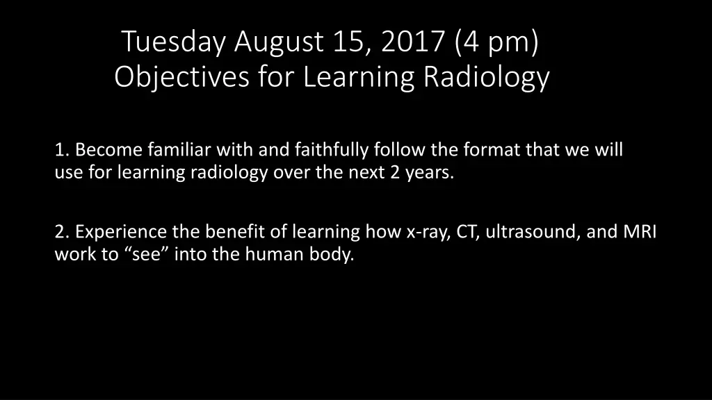
Tuesday August 15, 2017 (4 pm) Objectives for Learning Radiology 1. Become familiar with and faithfully follow the format that we will use for learning radiology over the next 2 years. 2. Experience the benefit of learning how x-ray, CT, ultrasound, and MRI work to see into the human body.
Objective 1: Format for learning radiology Please prepare by reviewing the pre-recorded information. Please come to class and actively participate. Don t let your knowledge/skills fade.
Objective 2 Understanding how an imaging modality works is better than memorizing patterns. Please evaluate the following images. You may not recognize the findings today, but you will.
The arrow points to the gallbladder. What is unusual (abnormal) about this gallbladder?
The arrow points to an object visible as which of the 5 basic densities? Diagnosis?
This is an ultrasound of the left breast. The arrow points to a mass in the breast. How would you advise this patient based on your findings?
1. What type of an imaging study is this (x-ray, CT, Ultrasound, MRI)? 2. What organ does the white arrow point to? 3. The white areas within this organ reflect which of the 5 basic radiographic densities? 4. What is your diagnosis?
The arrow points to the area of the pineal gland in the brain. 1. What kind of an imaging study is this? 2. Is this T1 or T2? 3. The bright white area is an area of: A. White matter B. Gray matter C. Fluid
B C A Two of these x-ray studies demonstrate a fracture. Which one does not?
What should you remember? 1. To be successful learning how to properly order and interpret medical imaging tests: Always review the pre-recorded information before class. Always come to class. Always participate in class. 2. If you understand how x-ray, CT, MRI, and ultrasound work to create an image, you will be able to choose the best imaging test for your patient and you will be able to recognize common pathologies on these medical images.


