Left Bundle Branch Block and its Impact
Detailed overview of Left Bundle Branch Block (LBBB) and its significance in cardiac health. Discusses diagnostic criteria, incidence rates, association with acute coronary syndrome (ACS), challenges in diagnosing MI, and more.
Download Presentation

Please find below an Image/Link to download the presentation.
The content on the website is provided AS IS for your information and personal use only. It may not be sold, licensed, or shared on other websites without obtaining consent from the author.If you encounter any issues during the download, it is possible that the publisher has removed the file from their server.
You are allowed to download the files provided on this website for personal or commercial use, subject to the condition that they are used lawfully. All files are the property of their respective owners.
The content on the website is provided AS IS for your information and personal use only. It may not be sold, licensed, or shared on other websites without obtaining consent from the author.
E N D
Presentation Transcript
INTRODUCTION ~ 1 % (0.2 to 1.1 % in different populations) Incidence Increases with age Etiology Structural and functional Structural CAD ACS Cardiomyopathy SHTN Aortic valve disease Post cardiac surgery esp aortic root surgeries Functional Rate dependent BBB
DEFINATION Diagnostic criteria are defined by the American College of Cardiology (ACC) and American Heart Association (AHA) as follows: Rhythm must be of super-ventricular origin (EG: ventricular activation coming from atrial or AV nodal activation) QRS Duration greater than 120 ms Lead V1 should have either a QS or a small r wave with large S wave Lead V6 should have a notched R wave and no Q wave
1. QRS duration greater than or equal to 120 ms in adults, (>100 ms in children 4 to 16 years ,>90 ms in children less than 4 years) 2. Broad notched or slurred R wave in leads I, aVL, V5, and V6. 3. Absent q waves in leads I, V5, and V6. 4. R peak time greater than 60 ms in leads V5 and V6 5. ST and T waves usually opposite in direction to QRS. 6. Positive concordance may be normal. 7. Negative concordance are abnormal. 8. The appearance of LBBB may change the mean QRS axis in the frontal plane. AHA/ACCF/HRS Recommendations for the Standardization and Interpretation of the Electrocardiogram
LBBB & MI 2% of all patients who present with a suspected ACS ECG changes of LBBB can mask & mimics acute MI changes Not all new onset or presumed new onset LBBB is due to coronary artery disease More likely to be older, female, and have a history of pre-existing cardiovascular disease, hypertension, and congestive heart failure than non- BBB patients with ACS These patients are at increased risk of adverse outcomes Not due to LBBB but by underlying ischemic and structural heart disease that has contributed to LBBB
The diagnosis of MI in the setting of LBBB is especially challenging by ECG. Left ventricular activation is delayed in LBBB and the initial septal activation is from right to left (opposite of the normal situation), septal Q waves indicative of an MI are absent. Secondary ST-T wave abnormalities that occur in LBBB obscure the recognition of injury currents in ischemia and infarction.
2004 ACC/AHA STEMI guidelines -New or presumed new onset LBBB is considered STEMI equivalent. LBB more diffuse structure than RBB LBBB as part of MI indicates extensive myocardial damage ( very rarely discrete lesion in proximal conduction system). 2013 ACC/AHA STEMI guidelines Has to be considered as STEMI equivalent, but not in isolation. Sgarbossa criteria should be also considered ESC 2017 ECG diagnosis of STEMI difficult with LBBB. Concordant ST elevation best indicator of ongoing ischemia Clinical suspicion of ongoing Ischemia LBBB should be managed similar to STEMI patients Fourth Universal Definition of Myocardial Infarction (2018) --In patients with LBBB, ST-segment elevation 1 mm concordant with the QRS complex in any lead may be an indicator of acute myocardial ischemia.
SGARBOSSA CRITERIA Score of 3 has specificity for AMI greater than 95% and is associated with higher 30-day mortality compared with LBBB patients with discordant ST-segment elevation alone Sensitivity of a Sgarbossa score of 3 is only approximately 20%
MODIFIED SGARBOSSA CRITERIA A. 1 lead with 1 mm of concordant STelevation B. 1 lead of V1-V3 with 1 mm of concordant ST depression C. 1 lead anywhere with 1 mm STE and proportionally excessive discordant STE, as defined by 25% of the depth of the preceding S-wave. Any of above three is STEMI equivalent Improved sensitivity but with reduced specificity compared with the original Sgarbossa criteria (90 vs. 98%)
OTHER SIGNS OF MI IN LBBB The presence of QR complexes in leads I, V5, or V6 or in II, III, and avf with LBBB strongly suggests underlying infarction Prominent notching of the ascending part of a wide S wave in the midprecordial leads (V3 V4) - Cabrera's sign Prominent notching in ascending limb of a wide R wave in lead I, aVL, V5, or V6 - Chapman's sign
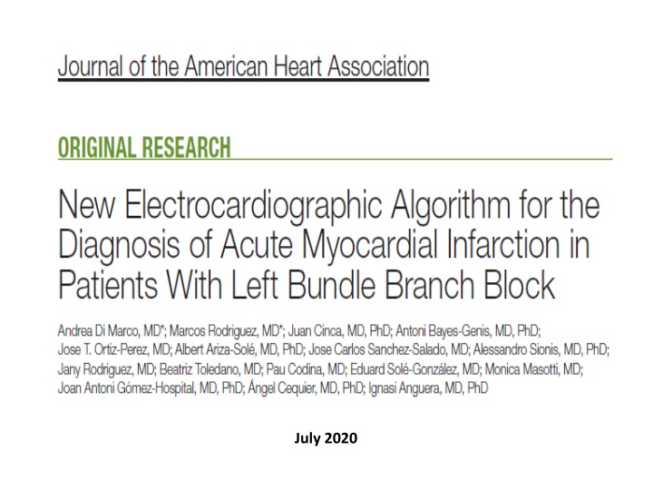
New Electrocardiographic Algorithm for the Diagnosis of Acute Myocardial Infarction in Patients With Left Bundle Branch Block
0BJECTIVE OF THIS STUDY The electrocardiographic diagnosis of AMI in the presence of LBBB improved by this BARCELONA algorithm.
Study Design and Patient Selection Retrospective, observational cohort study involving 4 referral hospitals for Ppci. Included all patients referred for pPCI because of new or presumed new LBBB. October 2009 and December 2014 As derivation sample, January 2015 until June 2018 - as validation sample. Control group - Patients with complete LBBB who attended the emergency department other than acute coronary syndrome or who were referred to this center for electrophysiological study or cardiac pacemaker implantation.
AMI was diagnosed in the presence of either an acute coronary artery occlusion (grade 0 of TIMI flow grading) or an acute coronary lesion with TIMI flow 1 associated with a troponin rise and fall above the 99th percentile upper reference limit. STEMI EQUVALENT Culprit artery occlution or acute non-occlusive coronary lesions with cardiac troponin I (cTnI) or cardiac troponin T (cTnT) ratios were 10 or when the creatine kinase MB isozyme (CK-MB) ratio was 5.
TOTAL - 484 patients Derivation cohort(163), Validation cohort (107) and Vontrol group (214) AMI diagnosed - 61(37%)derivation cohort and 40 (37%)validation sample.
Diagnostic Yielding of the ECG in the Entire Cohort of Patients Referred for pPCI The application of a Sgarbossa score 3 and the Modified Sgarbossa rules in 270 patients with LBBB referred for pPCI (101 diagnosed with AMI) would have missed 67 and 36 patients with AMI, respectively. By contrast, the BARCELONA algorithm would have missed only 6 patients. After pPCI, the BARCELONA algorithm became negative in 93% of patients with AMI who were positive before reperfusion.
CONTROL GROUP The BARCELONA algorithm was positive in 21 of 214 patients (10%), thus achieving a 90% specificity. 2 had ST elevation 1 mm (0.1 mV) concordant with QRS polarity, 1 had concordant ST depression 1 mm (0.1 mV), 17 had discordant ST deviation 1 mm (0.1 mV) in leads with max (R|S) voltage 6 mm (0.6 mV), and 1 had both ST-segment elevation 1 mm (0.1 mV) concordant with QRS polarity and discordant ST deviation 1 mm (0.1 mV) in leads with max (R|S) voltage 6 mm (0.6 mV). Thus, in this control group, the majority (81%) of false-positive Cases of the BARCELONA algorithm were attributable to the presence of discordant ST deviation 1 mm (0.1 mV) in leads with max (R|S) voltage 6 mm (0.6 mV).
CONCLUSIONS This study identified and validated the new ECG algorithm BARCELONA based on the presence of concordant ST deviation 1 mm (0.1 mV) in any ECG lead and/or discordant ST deviation 1 mm (0.1 mV) in leads with max (R|S) voltage 6 mm (0.6 mV). This algorithm significantly improved the diagnosis of AMI as compared with previous ECG rules, achieving a diagnostic performance for AMI similar to that of ECG in patients without LBBB.

![Guardians of Collection Enhancing Your Trading Card Experience with the Explorer Sleeve Bundle [4-pack]](/thumb/3698/guardians-of-collection-enhancing-your-trading-card-experience-with-the-explorer-sleeve-bundle-4-pack.jpg)


