Left lower quadrant abdominal pain and mass
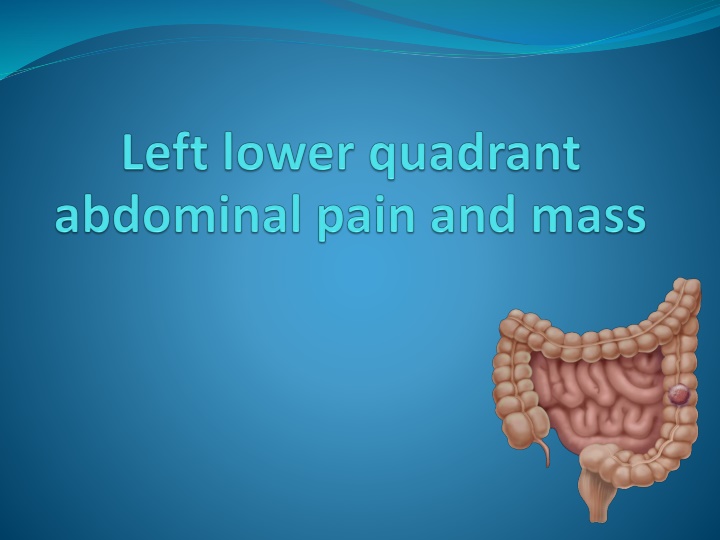
Various causes of masses in the left lower quadrant, including conditions affecting the skin, subcutaneous tissue, muscles, sigmoid colon, tube and ovary, lymph nodes, ilium, and more. It also covers causes of left lower quadrant pain and provides information on colorectal cancer and adenomatous polyps.

Download Presentation

Please find below an Image/Link to download the presentation.
The content on the website is provided AS IS for your information and personal use only. It may not be sold, licensed, or shared on other websites without obtaining consent from the author.If you encounter any issues during the download, it is possible that the publisher has removed the file from their server.
You are allowed to download the files provided on this website for personal or commercial use, subject to the condition that they are used lawfully. All files are the property of their respective owners.
The content on the website is provided AS IS for your information and personal use only. It may not be sold, licensed, or shared on other websites without obtaining consent from the author.
E N D
Presentation Transcript
left lower quadrant mass causes 1-Skin Sebaceous cyst (malformation) Abscess (inflammation) Primary and metastatic carcinomas (neoplasm) Contusion(truma) 2-Subcutaneous Tissue and Fascia Hernia Cellulitis Metastatic carcinoma Contusion Lipoma 3-Muscle Myositis Contusion
left lower quadrant mass causes 4-Sigmoid Colon Diverticulum Diverticulitis and abscess Carcinoma and polyp Perforation Volvulus Contusion Intestinal obstruction Tuberculosis Foreign body Granulomatous and ulcerative colitis
left lower quadrant mass causes 5-Tube and Ovary Hydatid cyst of Morgagni Tubo-ovarian abscess Ovarian cyst and carcinoma Ectopic pregnancy 6-Iliac Artery and Veins and Aorta Aneurysm Thrombophlebitis
left lower quadrant mass causes 7-Lymph Nodes Tuberculous and acute infectious adenitis Metastatic tumor 8-Ilium Osteomyelitis Sarcoma Fracture or contusion
Left lower quadrant pain causes 1-Abdominal abscess 2. Colonic volvulus 3. Constipation 4. Crohn's disease 5. Cystitis 6. Diverticular Disease 7. Ectopic pregnancy 8. Inguinal hernia 9. Intussusception 10. Ovarian cysts 11. Pelvic Inflammatory Disease 12. Pelvic abscess 13. Rectal abscess 14. Tuberculosis 15. Ulcerative colitis 16. Ulcerative proctosigmoiditis 17. Uterine fibroids
Colorectal cancer *Includes cancerous growths in the colon, rectum and appendix. *Precursor of ~90% of colorectal cancers is the adenomatous polyp. These mushroom-shaped growths are usually benign, but some develop into cancer over time. *Very curable if detected in early stage
colorectal cancers adenomatous polyp
Colorectal cancer *The transition from normal mucosa to polyp to invasive cancer is usually a lengthy process (7 12 years in many cases). Polyp size correlates to cancer probability Polyps < 1 cm 1% are cancerous Polyps > 2 cm 30% are cancerous
Human colon carcinogenesis progresses by the dysplasia/adenoma to carcinoma pathway
Epidemiology *Thereare nearly one million new cases of colorectal cancer diagnosedworld-wide each year and half a million deaths. *Most frequent form of canceramong persons aged 75 years and older. *It is the fifth most common form of cancer in the United States and the third leading cause of cancer-related death in the Western world.
Epidemiology 30% - 50% of population will develop adenomatous polyps over lifetime 1% - 3% of polyps become malignant Most remain asymptomatic & undetected Prevalence of polyps increases with age 50% of men, 40% of women by age 50 > 90% of CRC diagnosed after 55 yrs
pathology Colorectal cancer is a disease originating from the epithelial cells lining the colon or rectum , as a result of mutations along the 'Wnt signaling pathway. Some of the mutations are inherited, and others are acquired. The most commonly mutated gene in all colorectal cancer is the APC gene, which produces the APC protein.
Colon Cancer Module The malignant potential of a polyp based on histology is: tubular < tubulovillous < villous. Cancer risk also rises with increasing size of the polyp. The table below shows the incidence of invasive carcinoma related to polyp histology and size based on analysis of 7000 polypectomy specimens. Tubular Tubulovillous Villous size cm 0.5-0.9 2.5% 1.5% 0.3% 5.7% 6.4% 3.6% 1.0-1.9 17.0% 11.4% 6.5% 2.0-2.9 20.0% 16.0% 11.0% >3.0 pdf_logo Back Colonic polyps article Colonic polyps article Back Images from: www.endoatlas.com/atlas_co.html
Colon Cancer Module Hyperplastic Polyps www.GI-Pathology.net www.GI-Pathology.net Endoscopic image of hyperplastic polyps The epithelial cells at the base of the crypt (regenerative zone) have mildly enlarged, but uniform nuclei and brisk mitotic rate, feature which is normally present in reactive colonic mucosa Back
Colon Cancer Module Tubular Adenoma www.GI-Pathology.net www.endoatlas.com/atlas_co.html The adenomatous polyp has a smooth outline and is composed of numerous architecturally simple crypts with mild irregularity in size and shape Endoscopic image of a tubular adenoma Back
Colon Cancer Module Villous Adenoma www.GI-Pathology.net www.endoatlas.com/atlas_co.html Villous adenoma with glands that extend straight down from the surface to the base as fingerlike projections Endoscopic image of a villous adenoma Back
Colon Cancer Module Tubulovillous Adenoma www.GI-Pathology.net www.endoatlas.com/atlas_co.html Endoscopic image of a tubulovillous adenoma Tubulovillous adenoma with 80% tubular histology and 20% villous histology Back
The most common colon cancer cell type is adenocarcinoma which accounts for 95% of cases. Other, rarer types include lymphoma and squamous cell carcinoma.
Local symptoms *change in bowel habit *feeling of incomplete defecation (tenesmus) and reduction in diameter of stool rectal cancer. *passage of bright red blood in the stool. *mucus *Melena when the disease is located in the beginning of the large bowel.
Local symptoms Large tumor: *Bowel obstruction(constipation, abdominal pain, abdominal distension, vomiting, perforation and peritonitis) *Noticed by physical examination.
Local symptoms Local invasion: -blood or air in the urine (invasion of the bladder) -vaginal discharge (invasion of the female reproductive tract).
Constitutional symptoms *chronic occult bleeding cause iron deficiency anemia fatigue, palpitations and pallor. *decreased appetite and wieght loss. paraneoplasticsyndrome: *Fever(unusual) *thrombosis, usually deep vein thrombosis (most common)
Metastatic symptom Colorectal cancer most commonly spreads to the liver jaundice , abdominal pain.
Risk factor Average risk AGE: population > 50 years ~ 75% of CRC cases occur in this group *Before 50 uncommon unless a family history of early colon cancer.
Risk factor High risk Factors include personal and/or familial history of CRC or polyps, genetic syndromes (hereditary CRC), history of inflammatory bowel disease ~ 25% of CRC cases occur in this group
Risk factor Hereditary: Familial adenomatous polyposis (FAP) carries a near 100% risk of developing colorectal cancer by the age of 40 if untreated Hereditary nonpolyposis colorectal cancer (HNPCC) or Lynch syndrome
Risk factor Inflammatory bowel disease: About one percent of colorectal cancer patients have a history of chronic ulcerative colitis. Crohn'sdisease have a risk of colorectal cancer, but less than that of patients with ulcerative colitis.
Colon Cancer Module Inflammatory Bowel Disease Ulcerative Colitis & Crohn s Colitis www.learningradiology.com www.GI-Pathology.net www.GI-Pathology.net Crohn s Colitis endoscopic view Ulcerative Colitis gross specimen Back Barium enema: colon with "lead-pipe" appearance in ulcerative colitis Back
Familial Adenomatous Polyposis
Risk for Colorectal Cancer Dietary factors *High fat diets correlate w/ high rates of CRC *Protective effects of fiber are still unproven *Excessive calorie intake may enhance risk Behavioral factors *Physical activity may reduce risk *Excessive alcohol use and smoking have been associated with increased risk
Colorectal cancer risk *Women who have had cancer of the ovary, uterus, or breast are at higher risk of developing colorectal cancer. *Exogenous estrogens such as hormone replacement therapy (HRT), tamoxifen, or oral contraceptives might be associated with colorectal tumors *Lifestyle changes could decrease the risk of colorectal cancer as much as 60-80%.
Early Detection of Colorectal Cancer Detecting and removing polyps has been shown to reduce incidence of CRC Commonly used screening tests include Digital Rectal Exam Double-Contrast Barium Enema (DCBE) Fecal Occult Blood Test (FOBT) Flexible Sigmoidoscopy (FS) Colonoscopy
Digital Rectal Examination (DRE) *Not effective as CRC screening test *Sensitivity for CRC less than 10% *Often used to obtain stool sample for FOBT with chance for increased false positive results
Double-Contrast Barium Enema *Radiologic study barium is used as contrast material to visualize lumen of the colon *Effectiveness as screening test debated *National Polyp Study: Sensitivity of 48% for polyps > 1 cm *Further evaluation required if polyps detected *Less risk of perforation than endoscopic exams *May be recommended with flexible sigmoidoscopy for CRC screening
This is an x-ray called barium enema. The area in the circle shows an advanced cancer of colon that has produced an apple core lesion. The name apple core comes from the x-ray appearance of circumferentially eaten apple.
This represents the infiltration of the cancer into the muscular wall of the colon and the consequent loss of elasticity of the colon itself
Double-Contrast Barium Enema Rectal cancer Colon cancer
Fecal Occult Blood Testing (FOBT) *Detects blood from cancers or large polyps *Bleeding is intermittent and increases with polyp size and stage of cancer *Hemoccult II (guaiac-based) most widely used (0ne of FOBT type) *Inexpensive and easy to perform *Diet and medications affect results False positives: oral iron, aspirin, NSAIDs, anticoagualants False negatives: vitamin C *33% CRC mortality reduction with annual screening
Sigmoidoscopy Advantages Relatively accurate Quick procedure performed w/out sedation Inexpensive
Sigmoidoscopy Disadvantages *Misses 40% - 50% of CRC and polyps *Risk of colon perforation is 1 to 2 per 10,000 exa *Evidence for most effective screening interval is inconclusive
Colonoscopy *95% of CRC in reach of colonoscope *could eliminate 80% to 90% of CRC mortality in population over age 50 years *Diagnostic use after positive results on FOBT or FS *Recommended as initial screening test for high risk individuals
Polyps can be detected and removed during colonoscopy. Studies show this procedure would decrease by > 80% the risk of cancer death, started by the age of 50, and repeated every 5 or 10 years.
Colonoscopy Disadvantages Expensive Negative impact on patient s daily life Trained endoscopists must perform Bowel perforation most serious complication 1 to 3 per 1000 procedures
CRC Screening Guidelines Screening should be individualized according to age and comorbidities Three screening options suggested: *Double-contrast barium enema (DCBE) plus FS every 5 years *Colonoscopy every 10 years *FOBT annually and flexible sigmoidoscopy (FS) every 5 year.
Monitoring Carcinoembryonic antigen (CEA) is a protein found on virtually all colorectal tumors. CEA may be used to monitor and assess response to treatment in patients with metastatic disease. CEA can also be used to monitor recurrence in patients post- operatively.
staging TNM (for tumors/nodes/metastases) system "T" denotes the degree of invasion of the intestinal wall, "N" the degree of lymphatic node involvement, and "M" the degree of metastasis.
staging Dukes system A - Tumourconfined to the intestinal wall B - Tumour invading through the intestinal wall C - With lymph node(s) involvement (this is further subdivided into C1 lymph node involvement where the apical node is not involved and C2 where the apical lymph node is involved)


")


")