Locomotion and Movement in Living Beings
Movement is an essential feature of living organisms, with various forms observed in both animals and plants. Different types of movements, such as amoeboid, ciliary, and muscular, play crucial roles in locomotion and other bodily functions. Muscles, as specialized tissues, are key for movement and contraction, characterized by unique properties. Understanding the mechanism of muscle contraction sheds light on the intricate processes involved in movement.

Download Presentation

Please find below an Image/Link to download the presentation.
The content on the website is provided AS IS for your information and personal use only. It may not be sold, licensed, or shared on other websites without obtaining consent from the author.If you encounter any issues during the download, it is possible that the publisher has removed the file from their server.
You are allowed to download the files provided on this website for personal or commercial use, subject to the condition that they are used lawfully. All files are the property of their respective owners.
The content on the website is provided AS IS for your information and personal use only. It may not be sold, licensed, or shared on other websites without obtaining consent from the author.
E N D
Presentation Transcript
Chapter - 17 Locomotion and Movement
Movement is one of the significant features of living beings. Animals and plants exhibit a wide range of movements. Streaming of protoplasm in the unicellular organisms like Amoeba is a simple form of movement. Movement of cilia, flagella and tentacles are shown by many organisms. Human beings can move limbs, jaws, eyelids, tongue, etc. Some of the movements result in a change of place or location. Such voluntary movements are called locomotion. Walking, running, climbing, flying, swimming are all some forms of locomotory movements
TYPES OF MOVEMENT Amoeboid-macrophages and leucocytes in blood exhibit amoeboid movement. It is effected by pseudopodia formed by the streaming of protoplasm (as in Amoeba). Ciliary - Ciliary movement occurs in most of our internal tubular organs which are lined by ciliated epithelium. Passage of ova through the female reproductive tract is also facilitated by the ciliary movement. Muscular- Movement of our limbs, jaws, tongue, etc, require muscular movement.
MUSCLE Muscle is a specialised tissue of mesodermal origin. About 40-50 per cent of the body weight of a human adult is contributed by muscles. They have special properties like excitability, contractility, extensibility and elasticity. Based on their location, three types of muscles are identified : (i) Skeletal (ii) Visceral and (iii) Cardiac
Mechanism of Muscle Contraction Each organised skeletal muscle in our body is made of a number of muscle bundles or fascicles held together by a common collagenous connective tissue layer called fascia. Each muscle bundle contains a number of muscle fibres. muscle fibre is lined by the plasma membrane called sarcolemma enclosing the sarcoplasm. Muscle fibre is a syncitium as the sarcoplasm contains many nuclei. The endoplasmic reticulum, i.e., sarcoplasmic reticulum of the muscle fibres is the store house of calcium ions A characteristic feature of the muscle fibre is the presence of a large number of parallelly arranged filaments in the sarcoplasm called myofilaments or myofibrils. Each myofibril has alternate dark and light bands on it. A detailed study of the myofibril has established that the striated appearance is due to the distribution pattern of two important proteins Actin and Myosin.
The light bands contain actin and is called I-band or Isotropic band, whereas the dark band called A or Anisotropic band contains muscle fibre is lined by the plasma membrane called sarcolemma enclosing the sarcoplasm. Muscle fibre is a syncitium as the sarcoplasm contains many nuclei. The endoplasmic reticulum, i.e., sarcoplasmic reticulum of the muscle fibres is the store house of calcium ions. Both the proteins are arranged as rod-like structures, parallel to each other and also to the longitudinal axis of the myofibrils.
Structure of Contractile Proteins Each actin (thin) filament is made of two F (filamentous) actins helically wound to each other. Each F actin is a polymer of monomeric G (Globular) actins. Two filaments of another protein, tropomyosin also run close to the F actins throughout its length. A complex protein Troponin is distributed at regular intervals on the tropomyosin. In the resting state a subunit of troponin masks the active binding sites for myosin on the actin filaments Each myosin (thick) filament is also a polymerised protein. Many monomeric proteins called Meromyosins (Figure 20.3b) constitute one thick filament. Each meromyosin has two important parts, a globular head with a short arm and a tail, the former being called the heavy meromyosin (HMM) and the latter, the light meromyosin (LMM).
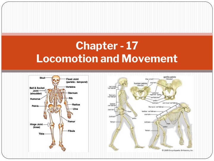
SKELETAL SYSTEM Skeletal system consists of a framework of bones and a few cartilages It is grouped into two principal divisions the axial and the appendicular skeleton. Axial Skull 28 Cranium 8,Facial 14,Ear Oscicle 6 Vertibral Column 27 Hyiod bone 1,Vertibrae(in Adult) 26 Tharax 25- Sternum 1,ribs 24 Appendicular Skeleton Upper region Pectoral girdle 4,arm and hands 60 Lower Region Pelvic girdle 2,Legs and feets 60 Total Bones - 206
Joint are essential for all types of movements involving the bony parts of the body Fibrous joints do not allow any movement. This type of joint is shown by the flat skull bones which fuse end- to-end with the help of dense fibrous connective tissues in the form of sutures, to form the cranium. In cartilaginous joints, the bones involved are joined together with the help of cartilages. The joint between the adjacent vertebrae in the vertebral column is of this pattern and it permits limited movements.
Synovial joints are characterised by the presence of a fluid filled synovial cavity between the articulating surfaces of the two bones. Such an arragement allows considerable movement. These joints help in locomotion and many other movements. Ball and socket joint (between humerus and pectoral girdle), Hinge joint (knee joint), Pivot joint (between atlas and axis), Gliding joint (between the carpals) and Saddle joint (between carpal and metacarpal of thumb) are some examples.
DISORDERS OF MUSCULAR AND SKELETAL SYSTEM Myasthenia gravis: Auto immune disorder affecting neuromuscular junction leading to fatigue, weakening and paralysis of skeletal muscle. Muscular dystrophy: Progressive degeneration of skeletal muscle mostly due to genetic disorder. Tetany: Rapid spasms (wild contractions) in muscle due to low Ca++in body fluid. Arthritis: Inflammation of joints. Osteoporosis: Age-related disorder characterised by decreased bone mass and increased chances of fractures. Decreased levels of estrogen is a common cause. Gout: Inflammation of joints due to accumulation of uric acid crystals.