Lumbar Plexus and Associated Nerve Formation
The lumbar plexus of nerves is formed in the psoas major muscle, giving rise to various nerves that play crucial roles in innervating different regions of the body. Understanding the structure and function of the lumbar plexus is essential in grasping the complexities of nerve supply in the abdominal and pelvic regions.

Download Presentation

Please find below an Image/Link to download the presentation.
The content on the website is provided AS IS for your information and personal use only. It may not be sold, licensed, or shared on other websites without obtaining consent from the author.If you encounter any issues during the download, it is possible that the publisher has removed the file from their server.
You are allowed to download the files provided on this website for personal or commercial use, subject to the condition that they are used lawfully. All files are the property of their respective owners.
The content on the website is provided AS IS for your information and personal use only. It may not be sold, licensed, or shared on other websites without obtaining consent from the author.
E N D
Presentation Transcript
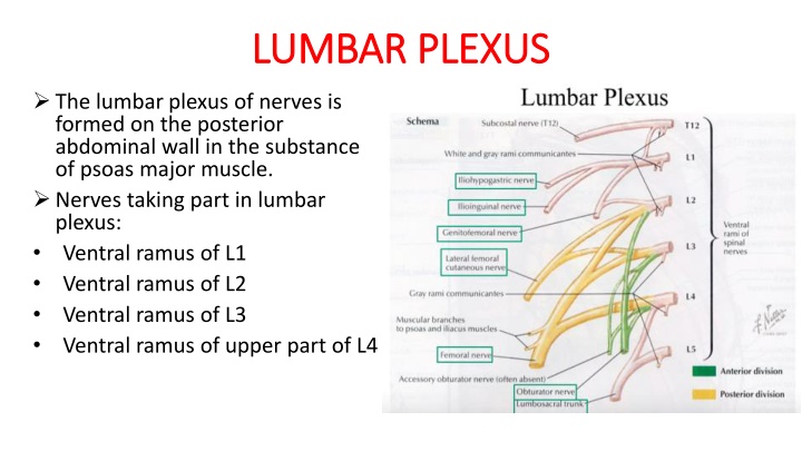
LUMBAR PLEXUS LUMBAR PLEXUS The lumbar plexus of nerves is formed on the posterior abdominal wall in the substance of psoas major muscle. Nerves taking part in lumbar plexus: Ventral ramus of L1 Ventral ramus of L2 Ventral ramus of L3 Ventral ramus of upper part of L4
CONT. CONT. Lower part of the fourth and the fifth ventral rami unite to form the lumbosacral trunk. Because the ventral ramus of fourth lumbar nerve bifurcates to take part in the lumbar plexus and the lumbosacral trunk,it is called the nervus furcalis. Formation of lumbar plexus- Ventral ramus of the L1 divides in to larger upper branch and smaller lower branch.the upper branch bifurcates in to iliohypogastric nerves(L1) and ilioinguinal nerve(L1). Lower branch unites with a branch from the second lumbar ventral ramus to form the genitofemoral nerve(L1,L2).
CONT. CONT. Second,third and fourth ventral rami divide in to dorsal and ventral divisions. Dorsal divisions of second and third give off smaller branches,which unites to form the lateral cutaneous nerve of thigh(L2,L3). Remaining parts of dorsal divisions of second,third and the entire dorsal division of the fourth unites to form femoral nerve(L2,L3 and L4). Ventral divisions of L2,L3 and L4 unite to form obturator nerve(L2,L3 and L4). Accessory obturator nerve,if present,receives fibres from third and fourth ventral division.
ILIOHYPOGASTRIC AND ILIOINGUINAL NERVE ILIOHYPOGASTRIC AND ILIOINGUINAL NERVE The iliohypogastric and ilioinguinal nerve(L1) come out of the lateral margin of psoas major muscle and then descend laterally on the front of quadratus lumborum. They enter the neurovascular plane of the anterior abdominal wall at the lateral border of quadratus lumborum muscle.
GENITOFEMORAL NERVE GENITOFEMORAL NERVE The genitofemoral nerve(L1,L2) emerges from the anterior surface of the psoas major muscle.it pierces the psoas sheath and then ends by dividing in to genital and femoral branches at a variable distance above the inguinal ligament. The genital branch enters the deep inguinal ring and traverses the inguinal canal to come out through the superficial inguinal ring.it supply the cremastor muscle and is sensory to scrotal skin in male and skin of mons pubis in female. The femoral branch dscends posterior to the inguinal ligament and enters the femoral sheath lateral to the femoral artery.it pierces the anterior wall of the femoral sheath and fascia lata to supply the skin of the upper part of femoral triangle.
LATERAL CUTANEOUS NERVE OF THIGH LATERAL CUTANEOUS NERVE OF THIGH The lateral cutaneous nerve of thigh(L2,L3) is also known as the lateral femoral cutaneous nerve. It emerges from the lateral margin of psoas major muscle and descends on the iliacus muscle. In the iliac fossa it is posterior to the lower part of descending colon on left side and cecum on the right side. It enters the thigh by passing behind or through the lateral end of inguinal ligament.at this site the nerve is vulnerable to compression,which may give rise to a condition called meralgia parasthetica.this nerve supplies anterolateral surface of thigh.
OBTURATOR NERVE OBTURATOR NERVE The obturator nerve(L2,L3,L4) takes origin from the lumbar plexus from the ventral divisions of ventral rami of lumbar second to fourth spinal nerves. It has an abdominal and intra pelvic course before it reaches the obturator foramen, through which it enters the medial compartment of thigh. Abdominal course-the obturator nerve emerges from thial border of psoas major muscle and enters the true pelvis behind the common iliac vessels and infront of the sacroiliac joint.
obturator nerve cont. obturator nerve cont. Intrapelvic course-The obturator nerve travels forwards on the medial surface of the obturator internus along the lateral wall of the true pelvis.the obturator vessels accompany the obturator nerve lying below it. The relation of obturator nerve to the ovarian fossa in female is important.the lateral surface of the ovary is in contact with the parietal peritoneum of ovarian fossa . The obturator nerve and vessels are retroperitoneal structures in this location. the inflammation of ovary irritates the obturator nerve.the patientexperiencespain along the medial side of thigh and knee.
OBTURATOR NERVE CONT. OBTURATOR NERVE CONT. Exit from pelvis-At the obturator foramen the obturator nerve divides in to anterior and posterior divisions,which along with obturator vessels enter the medial compartment of thigh. Accessory obturator nerve-in some subject accessory nerve(L2,L3) is present. It descends along the medial margin of psoas major muscle and follows the external iliac vessels.it passes behind the iguinal ligament to enter the thigh and supplies the pectineus and the hip joint.
FEMORAL NERVE FEMORAL NERVE The femoral nerve (L2,L3,L4) emerges from the lateral border of psoas major a little distance above the inguinal ligament. At this position it is located between the iliacus laterally and psoas major medially. It supplies branches to iliacus muscle and enters the femoral triangle ehind the inguinal ligament.