Luxations of Shoulder
Luxations of the shoulder joint in dogs, including the unique propensity in toy poodles and shelties. Learn about clinical signs, diagnosis methods like stress radiography, treatment options, and situations affecting successful reduction. Explore images and key anatomical points.

Download Presentation

Please find below an Image/Link to download the presentation.
The content on the website is provided AS IS for your information and personal use only. It may not be sold, licensed, or shared on other websites without obtaining consent from the author.If you encounter any issues during the download, it is possible that the publisher has removed the file from their server.
You are allowed to download the files provided on this website for personal or commercial use, subject to the condition that they are used lawfully. All files are the property of their respective owners.
The content on the website is provided AS IS for your information and personal use only. It may not be sold, licensed, or shared on other websites without obtaining consent from the author.
E N D
Presentation Transcript
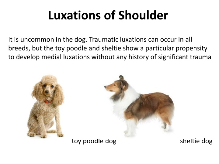
Luxations of Shoulder It is uncommon in the dog. Traumatic luxations can occur in all breeds, but the toy poodle and sheltie show a particular propensity to develop medial luxations without any history of significant trauma toy poodle dog sheltie dog
Clinical signs: With medial luxation the leg is usually carried with the elbow flexed and adducted and the lower limb abducted and supinated. With lateral luxation the position is similar except that the lower limb is adducted.
Superaglenoid tubercle Acromion process Greater tubercle Infraglenoid tubercle Intertubercular groove On palpation, the relative positions of the acromial process and the greater tubercle are the keys to determining the position of the humeral head relative to the glenoid. These points should be palpated on the normal limb and then compared with the affected limb.
Diagnosis: Clinical signs physical examination Stress radiography has been suggested as an objective method of measuring instability in this joint. Stress radiography : put a stress either (patient s own weight, force, or extra weight to carry) on specific organs usually joints
Situations that reduce the probability of a successful reduction: 1. 2. Congenital luxations discovered are usually irreducible because of severe malformations of both the glenoid and the humeral head. The presence of a severely eroded glenoid resulting from chronic luxation the presence of a dysplastic glenoid or humeral head. Treatment: 1. closed reduction and immobilization of the limb for approximately 2 weeks. 2. surgical treatment (if the joint remains unstable after reduction or if the luxation recurs.
Medial luxation of the left shoulder (ventrodorsal view) Lateral luxation of the left shoulder (ventrodorsal view). It occurs most often in large size breeds after trauma.
Excision Arthroplasty In case of the glenohumeral joint cannot be reconstructed adequately. Gunshot wounds occasionally damage the articular surfaces in such a way that nothing resembling normal joint function can result
Arthrodesis of Shoulder Joint Surgical fusion of the shoulder joint results in remarkably little functional disability because of the extreme mobility of the scapula. Arthrodesis: means the surgical fusion of a joint. In other words, the bones forming the joint are permanently joined together. Arthrodesis is a salvage procedure that is generally only performed when there are no other options to save the function of the joint.
Common indications for arthrodesis of the shoulder are 1. comminuted fractures of the glenoid, neck of the scapula, or head of the humerus. 2. Chronic shoulder luxations often result in severe erosion of the glenoid and humeral head, making surgical repair impossible. 3. Severe DJD (Degenerative joint disease )is a legitimate but uncommon indication. a last-resort salvage procedure