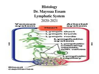
Lymphatic Organs and Tissue
Lymphatic organs play a crucial role in defending the body against foreign substances and maintaining fluid balance. Lymphatic tissue, composed of reticular fibers and various cells, is integral to the immune system. Lymphocytes, such as T-lymphocytes and B-lymphocytes, are key components responsible for immunity. This detailed overview covers the functions and composition of lymphatic organs and tissues.

Download Presentation

Please find below an Image/Link to download the presentation.
The content on the website is provided AS IS for your information and personal use only. It may not be sold, licensed, or shared on other websites without obtaining consent from the author.If you encounter any issues during the download, it is possible that the publisher has removed the file from their server.
You are allowed to download the files provided on this website for personal or commercial use, subject to the condition that they are used lawfully. All files are the property of their respective owners.
The content on the website is provided AS IS for your information and personal use only. It may not be sold, licensed, or shared on other websites without obtaining consent from the author.
E N D
Presentation Transcript
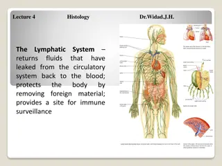
The main function of lymphatic organs are : 1-defence:protect the body against foreign substances and bacteria. 2-fluid balance :lymphatic organs maintain the fluid balance in the tissue which is called lymph ..which is the fluid that collecting from body tissue and return back to the blood circulation. The composition of the lymph is (ions, water, proteins, enzymes, cells such as lymphocyte).
Lymphatic Tissue The tissue in lymphatic organs which is composed of : _Reticular tissue: which is reticular fibers as network of reticularcells. _Free cells: are macrophage, plasma cells, and many lymphocyte. These are the main cells in immune system, also there is a cell antigen presenting cells cells are (interdigitating cell) or (dendirietic cells).
Lymphocytes _About 20% of the leukocytes in circulation. _They are functional unit of immune system. _These cells can recognize foreign antigen, they can distinguish self from non-self by their surface antigen receptors. _Types of lymphocytes: 1-T-lymphocyte 2-B-lymphocyte
1_T Lymphocyte _About 75% of circulating lymphocytes, (0-15) mm diameter. _Their stem cells originate in bone marrow as immature and undifferentiated, they complete their maturation in thymus, proliferate as lymphocytes recognize (thymus-depended lymphocytes). as T They are carried through blood stream to the spleen, lymph node and responsible immunity. for cellular
2_B lymphocyte -thymus in depended lymphocytes. -5-15% of the circulating lymphoid pool. -the main function of cells is the production of antigen- specific (immunoglobulin). -once activated B cell terminallydifferentiate in to plasma cells. -are originate and complete their maturation in bon marrow then reach (lymph node, circulation. spleen) through blood
They are responsible for humoral -immunity through which it reach with antigens that stimulate it to produce plasma cells that secret antibodies (immunoglobulin) (A, G, D, E, M) fluid. in the body _Lymphatic tissue are found in two forms: 1-Diffused lymphatic tissue. 2-Nodular lymphatic tissue.
_Diffused lymphatic tissue: Means loose aggregation of lymphatic tissue mainly lymphocyte, no special organization but as in filtration of lymphocyte, also lymphatic nodules no capsule. this type found in ct of lamina properia of digestive tract, respiratory passage tract. _Nodular lymphatic tissue: Dense aggregation of lymphatic lymphocyte as organized structure called nodules or follicles these follicles are about (0.2-1) mm in diameter, composed of zones: and urinary tissue mainly 2 areas or
1-Cenrtral area (germinal center)or(Secondary follicle): Appears as lightly stain area composed of larger sized lymphocyte or lymphoblast, have large cytoplasm and pale stained nucleus mitoticly active where there is antigenic stimulation and any few formed cells will be add to area. 2-Peripheral area (primary follicle): Appears as darkly stain lymphocyte with dark nucleus and little cytoplasm, these lymphocyte accumulated. peripheral composed of small sized are densely
_The lymphatic nodules is a variable either in size or position according to antigenic stimulation and they don t develop before birth. _Types of lymphoid tissue: 1-En Capsulated: contain capsule Spleen, Thymus, Lymph nodes. 2-Un en capsulated (or partiallyen capsulated) Tonsil, Peyer,s patches, Lymphoid nodules in digestive tract , respiratory tract, urinary and reproductive tracts.
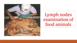
Lymph nodes Each lymph node is kidney shaped structure, various in size from (0.1-1.2) cm. Located along the course of major vessels in (neck, thorax, abdomen, axilla) to the filter the lymph. _Structure of lymph node: -oval shaped surrounded by adense fibrous c.tcapsule. -septaor trabeculeaextend from capsule in to lymph node and divided the node in to in completecompartments. -it has convex surface where afferent lymphatic vessels enter lymph node. -at concave side, found. -where blood vessels enter and leave lymph node, efferent lymphatic vessels leave hillum. hillum is the node through this
Structure of a Lymph Node Figure 20.4a, b
Histological structure Nodes have histological distinct regions: 1-cortex 2-Medulla 3-Paracortical area _Cortex Located beneath the capsule, composed of densely aggregation of lymphatic tissue, lymphatic nodules, some of them germinal center and some are diffuse lymphatic tissue may be found.
_Para cortical are Its located internal to the cortex, between cortex and medulla, composed of diffuse lymphatic tissue (T lymphocytes), macrophage and antigen-presenting cell (interdigitating cells). _Medulla It s the inner part of corex) composed of : A-Medullarycords Aggregate of cells as cords B lymphocyte cell and few T cell, plasma cells, macrophages are embedded in reticulartissue. lymph node (lighter than
B-Medullary sinuses Are lined by continuous endothelium in complete layer of reticular cells, macrophages supported by reticular fiber. Wall of sinuses allows free movement of lymphocytes between its endothelial cells.
Filteration of lymph through of lymph nodes: Start from afferent lymphatic vessels found in capsule of lymph nodes there are two capsular sinuses: 1.sub capsular sinuses 2.trabecular sinuses -subcapsularsinuses..located beneath the capsule. -the lymph run to trabecular sinuses and run with trabeculae in the cortex to medullary sinuses in medulla and then leave the lymph node through the hillum byefferent lymphaticvessels.
Afferent lymphatic vessels Sub capsular sinuses Trabicular sinuses Cortex Medulary sinuses Efferent lymphatic vessels Thoracic duct Blood stream Return to art
Function of lymph nodes: 1-filtration of lymph 2-defence response, lymphocyte mechanism through through T-lymphocyte phagocyte the immune and B- of , macrophage. 3-production of new lymphocyte through germinal center.