Malaria: Causes, Species, and Life Cycle
Malaria is a complex disease caused by protozoan parasites of the genus Plasmodium. Learn about the different species, their impact on human health, and the intricate life cycle involving both humans and mosquitoes.

Download Presentation

Please find below an Image/Link to download the presentation.
The content on the website is provided AS IS for your information and personal use only. It may not be sold, licensed, or shared on other websites without obtaining consent from the author.If you encounter any issues during the download, it is possible that the publisher has removed the file from their server.
You are allowed to download the files provided on this website for personal or commercial use, subject to the condition that they are used lawfully. All files are the property of their respective owners.
The content on the website is provided AS IS for your information and personal use only. It may not be sold, licensed, or shared on other websites without obtaining consent from the author.
E N D
Presentation Transcript
Anti-malarial drugs Mrs juliet olayinka
Malaria is a complex disease caused by protozoan parasites belonging to the genus Plasmodium. The human protozoa of the genus Plasmodium is classified thus; subkingdom: Protozoa; phylum: Apicomplexa; class: Sporozoasida; subclass: Coccidiasina; order: Eucoccidiorida; suborder: Haemospororina; family: Plasmodidae.
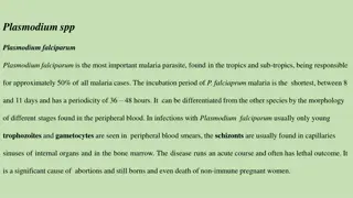
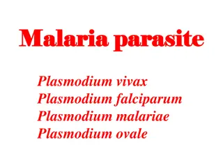
Five species of Plasmodia account for almost all human infections which are Plasmodium falciparum, Plasmodium vivax, Plasmodium malariae, Plasmodium ovale and Plasmdium knowlesi. Of these five species, Plasmodium falciparum is responsible for most severe disease and mortality and Plasmodium vivax accounts for most cases (although not most deaths) outside the Africa. Plasmodium knowlesi, a newly recognized 5thspecies is important in a small geographical range in Oceania.
LIFE CYCLE OF MALARIA The human malaria parasites undergo the following characteristics of life cycle. They undergo one cycle of the asexual division in the tissues called the exo-erythrocytic schizogony and another cycle of pigment producing asexual division in the red blood cells called the erythrocytic schizogony (in the vertebrate host). They also undergo a sporogonic development or cycle called (sporogony) in the body of the mosquito. Humans and other vertebrates act as the intermediate host for the parasite while the mosquito in which the sexual reproduction takes place is considered to be the definite one.
The Exoerythrocytic Stages of Malaria Parasites in Humans Malaria infection in the human host starts when the sporozoites are injected into the blood stream during a blood meal by an infectious mosquito. Sporozoites rapidly enter the circulation and localize to hepatocytes, where they transform, multiply, and develop into tissue schizonts. This primary asymptomatic stage (pre-erythrocytic or exoerythrocytic) lasts for 5 15 days, depending on the Plasmodium species. Tissue schizonts then rupture, releasing merozoites that enter the circulation, invade erythrocytes, and initiate the erythrocytic cycle
The liver trophozoite initially appears as a mononucleated round body into the cytoplasm of the host cells; subsequently it begins to develop and mutually asexually, then a mature schizont (the multinucleated stage of the parasite) is formed, then finally a large number of merozoites are released. The mature schizont is 30-70um large, it has no pigment (that there is no hemoglobin into the hepatocyte) and occupies the entire cell cytoplasm. The length of the schizogonic liver cycle is constant for each plasmodium species. The duration of this stage influences the length of the incubation period. It is usually short in Plasmodium falciparum (5.5-7 days), Plasmodium vivax (6-8days), Plasmodium ovale (9days) and Plasmodium malariae (13-16days)
Once the tissue schizonts burst in P. falciparum and P. malariae infections, no parasites remain in the liver. In P. vivax and P. ovale infections, tissue parasites (hypnozoites) persist and can produce relapses years after the primary attack. The liver trophozoites remain in the liver in a latent (dormant) stage for varying periods of time and are called hypnozoites.
The Erythrocytic Stage The erythrocytic stage is the stage causing the complex and varying spectrum of symptoms characterising the disease in humans. The blood phase of the life cycle is initiated when the merozoites from the liver schizonts are discharged into the circulation. The merozoite is 1nm in diameter consisting of a single nucleus and an adjacent cytoplasm
It immediately invades the erythrocyte and then enters the trophozoite stage. In erythrocytes, most parasites undergo asexual development from young forms to trophozoites and finally to mature schizonts. Schizont-containing erythrocytes rupture, producing febrile clinical attacks. The merozoites that are discharged into the circulation invade new erythrocytes to repeat the schizogonic cycle until the process is inhibited by the specific immune response or by chemotherapy.
The time required to complete the erythrocytic cycle is fixed among the parasite species. P. falciparum and P. vivax have a 48 hours development; P.ovale lasts for 50 hours while P.malariae has a longer cycle of 72 hours. Theoretically, the periodicity of the erythrocytic cycle would determine the classical cyclical presentation of symptoms
The Sporogonic Cycle In The Mosquito In the course of the schizogonic cycle, some of the merozoites become differentiated into sexual forms (the gametocytes); Two types of gametocytes are found in the peripheral blood- The female macrogamete the male micro-gametocytes. The male gametocyte has a nuclear material that is dispersed ( Prepairing to exflagellate) while the nuclear material of the female gametocyte is condensed.
The sporogonic cycle is initiated when mature female and male gametocytes are ingested by a suitable species of anopheles mosquito during a blood meal. Some erythrocytic parasites differentiate into sexual forms known as gametocytes. After a female mosquito ingests infected blood, exflagellation of the male gametocyte is followed by fertilization of the female gametocyte in the insect gut. The resulting zygote, which develops as an oocyst in the gut wall, eventually gives rise to sporozoites, which invade the salivary gland of the mosquito. The insect then can infect a human host by taking a blood meal.
Drugs used for prophylaxis treatment and prevention of relapse of malaria
Malaria is transmitted by the bite of infected femaleanopheles mosquitoes. During feeding, mosquitoes inject sporozoites, which circulate to the liver, and rapidly infect hepatocytes, causing asymptomatic liver infection (hepatic phase)(absent in falciparum; malariae) Merozoites released from the liver, rapidly infect erythrocytes to begin the asexual erythrocytic stage of infection that is responsible for human disease Multiplerounds of erythrocytic development, with production of merozoites that invade additional erythrocytes, lead to large numbers of circulating parasites and clinical illness
Release of merozoites subsequent to rupture of erythrocytes causes the clinicalattack of malaria. Some erythrocytic parasites also develop into sexual gametocytes, which are infectious to mosquitoes, allowing completion of the life cycle and infection of others In P vivaxand P ovaleparasites also form dormant liver hypnozoites, which are not killed by most drugs, allowing subsequent relapses of illness after initial elimination of erythrocytic infections AQQQ 11Q1QAAQqa
Malaria Transmission Cycle Exo-erythrocytic (hepatic) Cycle: Sporozoites infect liver cells and develop into schizonts, which release merozoites into the blood Sporozoires injected into human host during blood meal Parasites mature in mosquito midgut and migrate to salivary glands Dormant liver stages (hypnozoites) of P. vivax and P. ovale HUMAN MOSQUITO Erythrocytic Cycle: Merozoites infect red blood cells to form schizonts Some merozoites differentiate into male or female gametocyctes Parasite undergoes sexual reproduction in the mosquito
Signs and symptoms Initial manifestation of malaria are non- specific and resembles to flu like symptoms. The presentation includes headache, fever, shivering, arthralgia, myalgia. The paroxysm which includes fever spikes, chills and rigors are classical for malaria
The typical paroxysmal attack comprises of three distinct stages: a) Coldstage- The onset is with lassitude, headache, nausea and chilly sensation followed by rigors. The stage lasts for - 1 hour b) Hotstage- The patient feels burning hot, the skin is hot and dry to touch. Headache is intense. Pulse rate is high. The stage lasts for 2-6 hours c) Sweatingstage- Fever comes down with profuse sweating. The pulse rate gets slower, patient feels relieved. The stage lasts 2-4 hours
These paroxysms have different frequencies in different species of malarial parasites In P. vivax and P. ovale after every 2 days- Tertian fever In P. malariae after every 3 days- Quartan fever While in P. falciparum it recurs in every 36-48 hours These paroxysmal attacks coincide with the release of successive broods of merozites into the blood stream.
Relapse Vs Recrudesence Depending upon the cause , recurrence can be classified either as recrudescence or relapse Recrudescence is when symptoms return after a symptoms free period. It is due to parasites surviving in the blood as a result of inadequate or ineffective treatment. Relapse is when symptoms reappear after the parasites have been eliminated from blood but persist as dormant hypnozites in liver cells. Relapse is common in P.ovale and P.vivax infection Recrudescence is commonly seen in P.falciparum
Classification 1) 4-Aminoquinolines Chloroquine 2) Quinoline methanol Mefloquine Amodiaquine 3) Cinchona alkaloid Quinine Quinidine 4) Biguanides Proguanil (Chloroguanide)
Diaminopyrimidines 8-Aminoquinoline Sulfonamides & sulfone Sulfadoxine Antibiotics Pyrimethamine Primaquine Tafenoquine Sulfamethopyrazine Dapsone Tetracyclins Doxycycline
Sesquiterpine lactones Artesunate Artemether Arteether Halofantrine Lumefantrine Amino alcohols Pyronaridine Naphthyridine Naphthoquinone Atovaquone
Tissue schizonticides That eliminate pre erythrocytic/exo-erythrocytic stages in liver Primaquine: 15 mg/kg/day X 2 weeks(hypno) Proguanil Doxycycline Erythrocytic schizonticides act on erythrocytic parasites
Gametocides kill gametocytes in blood and prevent transmission to mosquitoes Primaquine gametocidal for all species. 45 mg single dose Immediately after clinical cure Cuts down transmission to mosquito
Clinical cure Terminate the episode of malarial fever Radical cure eliminate both hepatic and erythrocytic stages Causal prophylaxis Suppressive propylaxis
Clinical Cure Erythrocytic schizonticide is used to terminate the episode of malarial fever High efficacy Low efficacy
High efficacy Low efficacy 1) Artemesinin 2) Chloroquine 3) Amodiaquine 4) Quinine 5) Mefloquine 6) Halofantrine 7) Lumifantrine 8) Atovaquone Proguanil Pyrimethamine Sulfonamides Tetracyclins Clindamycin
Radical cure Eliminates bothhepatic and erythrocytic stages Vivax & ovale Erythrocytic schizonticide + Tissue schizonticide CQ + primaquine
Chloroquine resistance Quinine + Doxycycline/clindamycin + Primaquine Artemesinin based combination therapy + Primaquine
Causal prophylaxis Pre-erythrocytic phase which is the cause of malarial infection and clinical attacks is the target for this purpose Primaquine is the causal prophylactic for all species of malaria
Supressive prophylaxis Schizonticides which suppress the erythrocytic phase and thus attacks of malarial fever can be used as prophylactics Clinical disease does not appear
Supressive prophylaxis Mefloquine Doxycycline
Supressive prophylaxis Mefloquine 250 mg weekly Starting week before travel & taken till 4 weeks after return from endemic area for CQ resistant P. falciparum
Supressive prophylaxis Doxycycline 100 mg daily Starting day before travel & taken till 4 weeks after return from endemic area for CQ resistant P. falciparum CI in pregnantwomen & children <8years of age
Supressive prophylaxis Pregnancy One dose each in second & third trimester 1 month gap Pyrimethamine(75 mg)+ sulphadoxine(1500mg) In areas with high P.f endemicity
Goal To prevent and treat clinical attack of malaria. To completely eradicate the parasite from the patient's body. To reduce the humanreservoir of infection - cut down transmission to mosquito.
CHLOROQUINE Rapidly acting erythrocytic schizontocide against all species of plasmodia including the senstive strains of P. falciparum Controls most clinical attacks in 1-2 days with disappearance of parasites from peripheral blood in 1-3 days. No effect on Pre-erythrocytic and exo-erythrocytic phases of the parasite does not prevent relapses in vivax and ovale malaria. Only for clinical cure.
Mechanism of action: It is activelyconcentrated by sensitiveintra- erythrocytic plasmodia by accumulating in the acidic vesicles of the parasite and weakly basic nature it raises the vesicular pH and thereby interferes with degradation of haemoglobin by parasiticlysosomes Polymerization of toxic haeme to nontoxic parasite pigment hemozoin is inhibited by formation of chloroquine-heme complex
Haeme itself or its complex with chloroquine then damages the plasmodial membranes. Clumping of pigment and changes in parasite membranes follow: death Other related anti-malarials like amodiaquine quinine, mefloquine, lumefantrine act in an analogous manner
Resistance Reduceduptake and transport of chloroquine to food vacuole of plasmodium.
Pharmacokinetics Oral Widely distributed & concentrated in tissues like liver, spleen, kidney, lungs (several hundred-fold), skin, leucocytes and some other tissues Its selective accumulation in retina is responsible for the oculartoxicity seen with prolonged use
metabolized by liver excreted in urine. The early plasma t1/2varies from 3-10 days. Because of tighttissuebinding, small amounts persist in the body for longer time.
Adverse Effects GI intolerance Nausea, vomiting, abdominal pain, headache, anorexia, malaise, and urticaria are common. Dosing after meals may reduce some adverse effects. The long-term administration of high doses of chloroquine for rheumatologic diseases can result in loss of vision due to retinal damage. Cornealdeposits may occur affect vision: reversible
Contraindications & Cautions Chloroquine can ppt attacks of seizures, psoriasis or porphyria Cautious use Liver damage Severe GI, neurological, retinal & haematological diseases Safe in pregnancy and for young children
Other actions E. histolytica & Giardia lambia Anti-inflammatory Local irritant Local anaesthetic (on injection) Weak smooth muscle relaxant Anti-histaminic Anti-arrythmic properties
Therapeutic Uses Chloroquine is the preferred drug for clinical cure of Vivax Ovale malariae + some sensitivefalciparum strains Causes rapid clearance of fever & Parasitaemia
Extraintestinal amoebiasis/Hepatic amoebiasis/Amoebic Liver Abscess Due to high liver concentrations, it may be used for ameobic abscesses that fail initial therapy with metronidazole. Rheumatoid arthritis Other uses: Discoid lupus erythematosus Lepra reaction Photogenic reactions Infectious mononucleosis