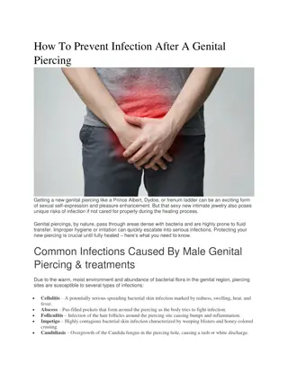
MALE EXTERNAL GENITAL ORGANS
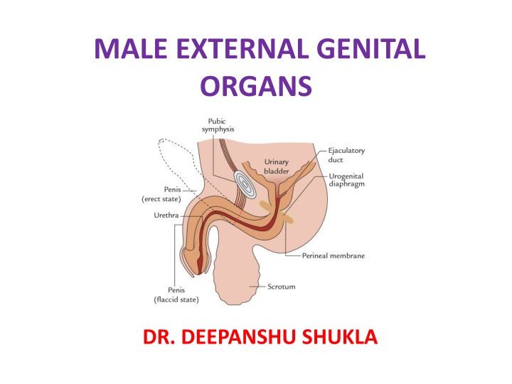
The male external genital organs consist of the penis, scrotum, testes, epididymis, and spermatic cords. The penis, essential for copulation, is divided into root (radix) and body (corpus) parts. The root comprises erectile tissue masses, while the body is further divided for its study, including the glans penis. Structural components like corpora cavernosa and corpus spongiosum play crucial roles during erection.

Download Presentation

Please find below an Image/Link to download the presentation.
The content on the website is provided AS IS for your information and personal use only. It may not be sold, licensed, or shared on other websites without obtaining consent from the author.If you encounter any issues during the download, it is possible that the publisher has removed the file from their server.
You are allowed to download the files provided on this website for personal or commercial use, subject to the condition that they are used lawfully. All files are the property of their respective owners.
The content on the website is provided AS IS for your information and personal use only. It may not be sold, licensed, or shared on other websites without obtaining consent from the author.
E N D
Presentation Transcript
MALE EXTERNAL GENITAL ORGANS DR. DEEPANSHU SHUKLA
INTRODUCTION The male genital organs are classified into two types external and internal. From surgical and clinical point of view, the male external genital organs include the following structures: 1. Penis. 2. Scrotum. 3. Testes. 4. Epididymis. 5. Spermatic cords.
PENIS The penis is the male organ of copulation and is traversed by the urethra, which provides passage for both urine and semen. PARTS= 1. Root or radix, an attached portion. 2. Body or corpus, a free pendulous portion.
Root (Radix) The root of the penis is situated in the superficial perineal pouch and is attached to the inferior aspect diaphragm. It comprises three masses of erectile tissue, viz. two crura (right and left) and the bulb of the penis. of the urogenital
Body (Corpus) The body of the penis is the free pendulous portion and lies in front of scrotum. It becomes continuous with the root of the penis. For convenience of understanding the body of penis is further divided into three parts: 1. body proper, 2. neck, and 3. glans penis study and proper
Body flaccid state, it is a long (cylindrical) structure, downwards and forwards. In erect state, it is directed upwards and forward and assumes prism-like shape on cross section with angles. Glans Penis= enlarged, conical structure at the distal end of the penis. Neck of Penis= It is an obliquely constriction the base of the glans. Proper= In the pendulous directed and a triangular rounded It is an grooved behind just
STRUCTURE It is composed of three elongated masses of erectile tissue, which considerable enlargement when engorged with blood during an erection. The three erectile tissue masses include: 1. two corpora cavernosa and 2. one corpus spongiosum are capable of
Corpora Cavernosa The two corpora cavernosa form the greater part of the body of penis. The two corpora cavernosa, throughout their length, lay in close apposition with one another and are surrounded by a common fibrous envelope called the tunica albuginea.
Transverse section through the body of the penis.
Corpus Spongiosum The corpus spongiosum is the forward continuation of the bulb. It is cylindrical and tapers slightly toward the distal end where expands to form a conical enlargement the glans penis. Throughout its length, it is traversed by the spongy part of the urethra. It is also surrounded by a thin fibrous tunica albuginea. it suddenly sheath of
COVERINGS Skin of Penis Prepuce of the skin Frenulum of the prepuce Preputial sac Superficial Fascia of the Penis
Skin of Penis It is remarkably thin, delicate, dark, and hairless. It envelops the body of penis completely. It is loosely attached to the fascial sheath of the penis and hence is freely mobile. At the neck of the penis, it is folded upon itself to form the prepuce or foreskin, which covers the glans for a variable distance.
Prepuce of the skin It is a fold of skin, which covers the glans for a variable extent and is attached to the neck of the penis.
Frenulum of the prepuce It is a median fold of skin on the ventral surface of glans, which passes from the inner surface of prepuce to the external urethral meatus. It is highly sensitive.
Preputial sac It is a potential space/cleft between the glans and the prepuce.
Superficial Fascia of the Penis It consists of two layers superficial and deep. The superficial layer is devoid of fat and consists of loose areolar tissue. It may contain few muscle fibres the peripenic muscle. The deep layer in the lower part of the anterior abdominal wall is condensed and forms the fascia of penis termed deep fascia of the penis or Buck s fascia. It surrounds both corpora cavernosa and corpus spongiosum, but does not extend beyond the neck of penis.
SUPPORTS OF PENIS The ligaments support the weight of the free pendulous part (body) of the penis. They are number: 1. Fundiform ligament 2. Suspensory ligament two in
ARTERIAL SUPPLY The penis is supplied by the following four pairs of arteries: 1. Deep arteries of the penis. 2. Dorsal arteries of the penis. 3. Arteries of the bulb. 4. Superficial dorsal arteries of penis.
VENOUS DRAINAGE 1. Superficial dorsal vein of the penis. 2. Deep dorsal vein of the penis.
NERVE SUPPLY 1. Sensory innervation: Dorsal nerve of penis. Ilioinguinal nerve. 2. Motor innervation: pudendal nerve. 3. Autonomic innervation: The autonomic nerves of the penis are derived from the pelvic (inferior hypogastric) plexus via the prostatic plexus. The sympathetic nerves parasympathetic fibres are vasodilator. The parasympathetic fibres (nervi erigentes) are derived from S2, S3, and S4 spinal segments. The autonomic fibres are distributed through the pudendal nerve. are vasoconstrictor while
Lymphatic Drainage Lymphatics from Glans- into deep inguinal nodes also called gland of Cloquet. Lymphatics from the rest of Penis- into the superficial inguinal lymph nodes.
Peyronies disease/chordee It occurs due to a thickening plaque corpora cavernosa, which expansion of a segment erectile during erection. As a result penis becomes curved especially during erection. localized or of the prevents of tissue
Phimosis It is a narrowing of the distal end of the (foreskin), prevents retraction over the glans penis may interfere with the micturition (passage of urine). prepuce which its and
Paraphimosis It is an uncommon condition in which narrowing prepuce insufficient interfere with the micturition, but the prepuce sufficiently tight to get stuck on the glans posteriorly on erection and thus interfere copulation. of the is to is just with
Circumcision It is the surgical removal of the prepuce (foreskin of the penis). In children and adults, the circumcision is sometimes required to relieve the patient from a tightly constricting prepuce (phimosis). The ritual of circumcision for religious reasons is one of the oldest operative procedures in the world.
SCROTUM The scrotum (L. scrotum = bag) is a large pendulous sac of skin located below and behind the penis. It is considered as an out-pouching of the lower part of the anterior abdominal wall. It contains the following structures: 1. Testes. 2. Epididymis. 3. Lower parts of the spermatic cords.
EXTERNAL FEATURES 1. The scrotum is divided into right and left halves by a median ridge or raphe, which indicates the line of fusion of the two halves of the scrotum. This ridge is continued forward in the midline to the undersurface of the penis and backward in the midline of the perineum to the anus. 2. The skin is rugose (corrugated) and dark in colour. The rugosity of the skin occurs due to the presence of subcutaneous dartos muscle. 3. The left half of the scrotum hangs lower than the right half, because the left spermatic cord is longer than the right spermatic cord.
LAYERS OF THE SCROTUM The scrotal wall from without made up following five layers: 1. Skin. 2. Dartos (which replaces the superficial fascia). 3. External spermatic fascia. 4. Cremasteric muscle and fascia. 5. Internal fascia. inward of is the muscle spermatic
Comparison between the layers of the anterior abdominal wall and the scrotum
BLOOD SUPPLY 1. Superficial external pudendal artery. 2. Deep external pudendal artery. 3. Scrotal branches of the internal pudendal artery. 4. Cremasteric artery, a branch of the inferior epigastric artery.
Scrotal Elephantiasis It is a clinical condition characterized by a massive swelling and enlargement of the scrotum accumulation interstitial the scrotal following blockage of lymph vessels slender worms filariasis (Wuchereria bancrofti). due to of in fluid wall by of


")
")