Mammary Glands
Discover the anatomy and function of mammary glands and breasts in the female reproductive system. Explore the structure, role of hormones, milk production, and blood supply in this comprehensive guide by Mrs. Mercy Deva Priya, Asst. Prof. Dept. of MHN.

Uploaded on | 1 Views
Download Presentation

Please find below an Image/Link to download the presentation.
The content on the website is provided AS IS for your information and personal use only. It may not be sold, licensed, or shared on other websites without obtaining consent from the author. If you encounter any issues during the download, it is possible that the publisher has removed the file from their server.
You are allowed to download the files provided on this website for personal or commercial use, subject to the condition that they are used lawfully. All files are the property of their respective owners.
The content on the website is provided AS IS for your information and personal use only. It may not be sold, licensed, or shared on other websites without obtaining consent from the author.
E N D
Presentation Transcript
Mammary Glands Mrs.Mercy Deva Priya Asst.Prof Dept of MHN
The breasts or mammary glands are accessory glands of the female reproductive system. They exist also in the male but in only a rudimentary form. During pregnancy these hormones stimulate further growth. After the baby is born the hormone prolactin from the anterior pituitary production of milk, and oxytocin from the posterior pituitary stimulates the release of milk in response to the stimulation of the nipple by the sucking baby, by a positive feedback mechanism. stimulates the
Structure of the breast The mammary glands consist of glandular tissue, fibrous tissue and fatty tissue. Each breast consists of about 20 lobes of glandular tissue, each lobe being made up of a number of lobules that radiate around the nipple. The lobules consist of a cluster of alveoli which open into small ducts and these unite to form large excretory lactiferous ducts. ducts, called
The lactiferous ducts converge towards the centre of the breast where they form dilatations or reservoirs for milk. Leading from each dilatation, or lactiferous sinus, is a narrow duct which opens on to the surface at the nipple. Fibrous tissue supports the glandular tissue and ducts, and fat covers the surface of the gland and is found between the lobes.
The nipple. This is a small conical eminence at the centre of the breast surrounded by a pigmented area, the areola. On the surface of the areola are numerous sebaceous glands (Montgomery's tubercles) which lubricate the nipple during lactation.
Blood supply, lymph drainage and nerve supply Arterial supplied branches of the axillary arteries and from the internal mammary and intercostal arteries. Venous drainage. anastomotic circle round the base of the nipple from which branches carry the venous blood to the circumference and end in the axillary and mammary veins. blood with supply. blood The breasts the are from thoracic This describes an
Lymph drainage. This is mainly into the axillary lymph vessels and nodes. Lymph may drain through the internal mammary nodes if the superficial route is obstructed. Nerve supply. The breasts are supplied by branches from the 4th, 5th and 6th thoracic nerves which contain sympathetic fibres. There are numerous somatic sensory nerve endings in the breast especially around the nipple. When these touch receptors are stimulated by sucking, impulses pass to the hypothalamus and the flow of the hormone oxytocin is increased, promoting the release of milk.
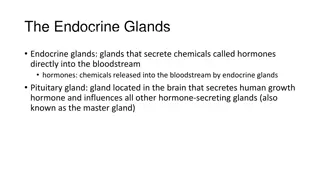
Function of the breast The mammary glands are only active during late pregnancy and after the birth of a baby when they produce milk (lactation). Lactation is stimulated by the hormone prolactin