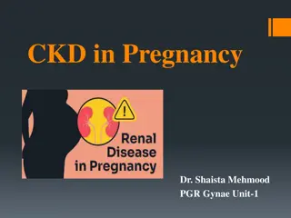
Managing Cardiovascular Pathology in Pregnant Women
Learn about the frequency and types of extragenital pathology in pregnant women, with a focus on cardiovascular issues. Find out how pregnant women with chronic diseases are monitored and treated throughout their pregnancy to ensure a safe outcome for both mother and baby.

Download Presentation

Please find below an Image/Link to download the presentation.
The content on the website is provided AS IS for your information and personal use only. It may not be sold, licensed, or shared on other websites without obtaining consent from the author. If you encounter any issues during the download, it is possible that the publisher has removed the file from their server.
You are allowed to download the files provided on this website for personal or commercial use, subject to the condition that they are used lawfully. All files are the property of their respective owners.
The content on the website is provided AS IS for your information and personal use only. It may not be sold, licensed, or shared on other websites without obtaining consent from the author.
E N D
Presentation Transcript
Frequency and types of extragenital pathology (EGP) at pregnant women. Now from 70 to 80% of pregnant women have chronic diseases and almost 90% of women during pregnancy have acute states (anemia, a pyelonephritis, etc.) which complicate its current. Main structure of EGP: 1. Pathology of cardiovascular system (the congenital and acquired heart diseases, arterial hypertension, hypotension, etc.). 2. Pathology of respiratory system (bronchitis, pneumonia, etc.). 3. Pathology of endocrine system (diabetes mellitus, pathology of a thyroid gland, etc.). 4. Digestive tract pathology (peptic ulcer, hepatitis, pancreatitis, etc.). 5. Pathology of an urinary system (cystitis, pyelonephritis, etc.). 6. Blood diseases (anemia, leukosis, etc.). 7. Infectious and parasitic diseases.
Chronic heart failure Activity of a rheumatic disease Pulmonary hypertensia 25 Risk HF Arrhythmia I isn't raised) F0 no no 40 II (are moderately raised) F1 I no > 40, but there is less systemic it is more than systemic Ciliary arrhythmia, etc. Ciliary arrhythmia, etc. III (high) F2a II The IV (extremely high) F2b, F3 III Tactics: I, II degrees of risk pregnancy aren't contraindicative; The III degree of risk pregnancy is contraindicative, is interrupted up to 12 weeks of a gestation; The IV degree of risk abortion according to vital indications in any term after achievement of clinical improvement of a state as a result of treatment is shown.
Maintaining pregnant women with cardiovascular pathology. Pregnancy is conducted in common by the cardiologist and the obstetrician- gynecologist. The obstetrician-gynecologist examines the pregnant woman at least 1 time in 2 weeks, the therapist at least 1 time in 3 weeks taking into account a condition of the patient. Planned stationary inspection and treatment of pregnant women with cardiovascular pathology is carried out 4 times during a gestation.
First hospitalization it is carried out up to 12 weeks or at the first address in/to the woman is hospitalized in therapeutic unit Purpose: specification of the arterial hypertension and solution of a question of opportunities of prolongation and features of conducting pregnancy or its discontinuing on medical indications. All doubts in the diagnosis or in a possibility of continuation of pregnancy are solved up to 12 weeks of a gestation.
Second hospitalization it is made in 18-20 weeks of pregnancy the woman is hospitalized in unit of pathology of pregnant women of maternity hospital Purpose: together with the therapist the possibility of prolongation of pregnancy is specified, control and correction of a hemodynamic is exercised, tactics of conducting pregnancy is discussed, if necessary preventive therapy is prescribed. The main medical and diagnostic objectives at this stage: 1. Assessment of a condition of the pregnant woman. 2. Assessment of the obstetric status and condition of a fetus. 3. Need of performing preventive treatment and correction of the taped disturbances.
Third hospitalization it is carried out to 28-30 weeks (the period of the maximum hemodynamic load) the woman is hospitalized in unit of pathology of pregnant women of maternity hospital Purpose: preventive, solves the same problems, as the second hospitalization.
Fourth hospitalization The hospitalization is carried out in 2-3 weeks prior to the estimated term of labors to a maternity hospital for the choice of a method of a delivery, antepartum preparation and correction of the taped disturbances in system. If there are indications, the patient is hospitalized at any gestational age: at activation of rheumatic process, emergence or intensifying of signs of a circulatory inefficiency, at any complications of pregnancy.
Delivery of women with heart diseases. Optimum delivery time at women with heart diseases is the term of a gestation of 37-38 weeks in case of a stable hemodynamic, lack of complications of a basic disease and favorable assessment of the obstetric status. Indications for an early delivery by natural birth canal or by Cesarean section: Aggravation of symptoms of the patient, weighting of a stage of CF, activity of rheumatic process, emergence of complications of a basic disease; lack of effect of treatment within 12-14 days; conditions that underlie obstetric indications for early delivery develop or aggravated.
Way of a delivery of patients with heart diseases: The I degree of risk vaginal delivery. Cesarean section, obstetric forceps are carried out only according to obstetric indications The II degree of risk - vaginal delivery + epiziotomyor perineotomy; Cesarean section at unstable parameters of a hemodynamics in the I period of labors. Obstetric forceps at unstable parameters of a hemodynamic in the II period of labors. The III degree of risk it is necessary to give Cesarean section, however preference to vagina labor with obligatory switching off of the II period of labors (obstetric forceps), with an anesthesiology grant. The IV degree of risk Cesarean section.
Management during labor PLACE OF INDUCTION: Most patients with cardiac disease go into spontaneous labor and deliver without any difficulty. However, induction (vaginal PGE2) may be employed in very selected cases for obstetric indications. One should guard against infection and pulmonary edema due to fluid overload.
Management during labor. First stage Position: The patient should be in lateral recumbent position to minimize aortocaval compression Oxygen is to be administrated (5-6 L/min) if required Analgesia in the majority, is best given by epidural Prophylactic antibiotics against bacterial endocarditis Fluids should not be infused more than 75 mL/hour to prevent pulmonary edema Careful watch of the pulse and respiration rate Cardiac monitoring and pulse oximetry can detect arrhythmias and hypoxemia early Central venous pressure monitoring may be needed in selected cases.
Prophylactic antibiotics for bacterial endocarditis Antibiotic prophylaxis during labor and 48 hours after delivery is considered appropriate. This is to prevent bacterial endocarditis. The recommended regimens include intravenous ampicillin 2 g and gentamicin 1.5 mg/kg (not to exceed 80 mg), at the onset or induction of labor followed by repeat doses 8 hours interval. High-risk patients are: a) Structural heart disease b) Rheumatic heart disease c) Cyanotic congenital heart disease d) Presence of dental and respiratory tract infections e) Hypertrophic cardiac myopathy f) Prosthetic heart valves g) Prior history of infective endocarditis h) Cardiac transplant
Management during labor. Second stage No maternal pushing and the tendency to delay in the second stage of labor is to be curtailed by forceps or ventouseunder pudendal and/or perineal block anesthesia. Ventouse is preferable to forceps as it can be applied without putting the patient in lithotomy position (raising the legs increases the cardiac load). Intravenous ergometrine with the delivery of the anterior shoulder should be withheld to prevent sudden overloading of the heart by the additional blood squeezed out from the uterus.
Management during labor. Third stage Conventional management is to be followed. Slight blood loss is not detrimental but if it is in excess, oxytocin can be given by infusion. This may be accompanied by aggressive diuresis by IV frusemide. It is better to administer oxytocin in preference to ergometrine in all cases of heart disease in third stage.
PLACE OF CESAREAN SECTION: In general, there is no indication of cesarean section for heart disease. CARDIAC INDICATIONS OF CESAREAN DELIVERY Coarctation of aorta Aortic dissection or aneurysm Aortopathywith aortic root > 4 cm In coarctation of aorta, elective cesarean section is indicated to prevent rupture of the aorta or mycotic cerebral aneurysm. The anesthesia should be given by expert anesthetist using either epidural (preferred) or general anesthesia.
PUERPERIUM The first days the bed rest, with extension is after the delivery prescribed at an unstable hemodynamics; In the early puerperal period the load is put on a stomach; Control of the ABP, diuresis, thermometry each 3-4 h; Antirheumaticdrugs, cardiotonics, antibiotics are prescribed; Anticoagulating therapy is carried out at valval prostheses, Eyzenmeyger'ssyndrome, primary pulmonary hypertension, chronic fibrillation of auricles, Cesarean section; Diuretic drugs according to indications; For 3-4 and 9-10 days it is after the delivery conducted complex cardiologic and laboratory researches (blood test, SRB, sialic acids, a coagulogram, an ECG, etc.). The extract of puerperas is carried out for 10-12 day after the delivery at satisfactory clinical laboratory data. Breastfeeding is not contraindicated unless there is failure. Anticoagulant therapy is not a contraindication of breastfeeding.
Congenital heart diseases: Atrial Septal Defect (ASD); Ventricular Septal Defect (VSD); Patent DuctusArteriosus (PDA); Fallot's tetralogy; Coarctation of aorta; Primary pulmonary hypertension; Stenosisof the valve of a pulmonary artery; Marfan's syndrome; Eisenmenger's syndrome.
The acquired heart diseases: mitral stenosis / failure; aortic stenosis / failure; mitral valve prolapse (MVP).
Contraindications to pregnancy at the acquired defects Pathology Relative contraindications Absolute contraindications Rheumatic carditis Latent Active (I, II St) Primary or recurrent rheumatic carditis In the last 24 months. In the last 12 months. FIIA, in the anamnesis repeated attacks of pulmonary asthma, disturbances of a circulation In the anamnesis of F1, attack of pulmonary asthma Mitral stenosis FIIB, atriomegaly, pulmonary hypertension The combined heart diseases F2 in the anamnesis, F1 episodes Kommisurotomy Effective Noneffective Infrequent and incontinuous attacks of a Bouveret's disease; full AV blockade (pulse >of 40 beats/min); frequent ventricular or supraventricular premature ventricular contraction; Ciliary arrhythmia, flutter, atrial fibrillation, full AV blockade (pulse <40 a beats/min), the progressing stenocardia Lesion of a myocardium, disturbance of a rhythm
Abortion: - in the presence of 1 absolute either 2 relative contraindications, - or 1 relative contraindication with two burdening factors (the chronic centers of an infection, an intolerance of heart and antirheumaticdrugs).
Contraindications to pregnancy at congenital defects the expressed aorta stenosis; coarctation of an aorta of 2-3 Art.; cyanosis forms of congenital defects (tetrad and Fallo's pentade, transposition of the main vessels, general arterial trunk, etc.); the stenosis of a pulmonary trunk which is followed by a right ventricular failure; a prolapseof the mitral valve with expressed by a prolapse of both cusps and a regurgitation; difficult disturbances of a rhythm; all congenital defects with a circulatory unefficiencyof FIIA, FIIB, FIII of stages.
Medical indications, for abortion in any term of a gestation: acute rheumatic fever; chronic rheumatic pericardis; illnesses (defects) of the mitral, aortal, three-leaved valve with a circulatory unefficiency; illnesses (defects) of mitral and aortal valves (combined), the illnesses which are characterized by the raised ABP; Ischemic heart disease; disturbance of a pulmonary circulation and heart failure, pulmonary heart; myocardites, perikardita; disturbances of a cordial rhythm (fibrillation and atrial flutter and ventricles), aortic aneurysm, embolism and thrombosis of arteries; disturbance of the blood circulatory system after the medical procedures.
ESSENTIAL HYPERTENSION IN PREGNANCY Apart from the specific hypertensive disorder in pregnancy (PIH), essential hypertension is the common hypertensive state in pregnancy. Its incidence varies from 1 to 3%.
DIAGNOSIS The diagnostic criteria are: 1. Rise of blood pressure to the extent of 140/90 mm Hg or more during pregnancy prior to the 20th week (molar pregnancy excluded), 2. Cardiac enlargement on chest radiograph and ECG, 3. Presence of medical disorders, 4. Prospective follow-up shows persistent rise of blood pressure even after 42 days following delivery. However, confusion in the diagnosis arises when the case is first seen in later months of pregnancy, especially when the prepregnant level of blood pressure remains unknown. Differential diagnosis with pre-eclampsia, gestational hypertension and essential hypertension are given below.
Differential diagnosis with pre-eclampsia, gestational hypertension and essential hypertension
EFFECTS OF PREGNANCY ON THE DISEASE: (1) There may be a midpregnancy fall of blood pressure in about 50%. However, the blood pressure tends to rise in the last trimester which may or may not reach its previous level, (2) In 50%, the blood pressure tends to rise progressively as pregnancy advances, (3) In about 20%, it is superimposed by pre-eclampsia evidenced by rise of blood pressure to the extent of 30 mm Hg systolic and 15 mm Hg diastolic associated with edema and/or proteinuria, (4) Rarely, malignant hypertension supervenes, (5) In 30%, there is permanent deterioration of the hypertension following delivery.
EFFECT OF THE DISEASE ON PREGNANCY: Maternal risk: In the milder form, the maternal risk remains unaltered but in the severe form or when superimposed by pre-eclampsia, the maternal and perinatal risk is much increased. Fetal risk: Due to chronic placental insufficiency, the babies are likely to be growth retarded. Preterm birth is high. In the milder form, with the blood pressure less than 160/100 mm Hg, the perinatal loss is about 10%. When blood pressure exceeds 160/100 mm Hg, the perinatal loss increases by three to four times and when complicated by pre-eclampsia, it increases further. Risk of placental abruption is high (0.5-10%).
MANAGEMENT: The principles of management are: (1) To stabilize the blood pressure to below 160/100 mm Hg, (2) To prevent superimposition of pre-eclampsia, (3) To monitor the maternal and fetal wellbeing, (4) To terminate the pregnancy at the optimal time. Preconceptional evaluation and counseling is essential to assess the etiology, severity of hypertension and possible outcome of pregnancy.
GENERAL MANAGEMENT: In mild cases with blood pressure less than 160/100 mm Hg, adequate rest (physical and mental), low-salt diet are all that are needed. The checkup should be more frequent 1-2 weeks interval up to 28 weeks and thereafter weekly. In severe cases or in cases of superimposed pre- eclampsia, the patients should be hospitalized and are placed in the treatment protocol as described under pre-eclampsia.
Medicamental therapy of arterial hypertension during pregnancy has to answer the following rules:drugs: 1. Considering the relative hypotension developing in the I trimester of pregnancy pressure during this period, as a rule, doesn't demand correction. 2. Purpose of drugs is recommended at the ABP> 150/100 mm Hg, and also at the ABP> 140/90 mm Hg in the presence of a hypertrophy of a left ventricle, a proteinuriaor symptoms of a preeclampsia. 3. Rising of the ABP more than 170/110 mm Hg demands urgent depression. 4. Anti-hypertensive drugs have to be effective and safe for a fetus.
For treatment of arterial hypertension at pregnant women are used now: Central -adrenomimetik (Methyldopum, Clophelinum). Beta adrenoblockers (metoprolol,bisoprolol, etc.). Use of an atenolol (selective beta1-adrenoblocker) is possible during the short period of time on late durations of gestation. At long use can lead to disturbance of fetal body height of a fetus. Blockers of calcium channels (Nifedipinum, amlodipin, etc.). Magnesium sulfate. APF inhibitors (captopril, enalapril) and blockers of receptors of angiotensin II are contraindicative to pregnant women since possess teratogenicaction. It is established that in II and III trimesters of pregnancy they are capable to oppress excretory function of kidneys of a fetus, to cause an oligoamnios.
Current of arterial hypertension during pregnancy: in the I trimester of pregnancy temporary depression of the ABP at patients with arterial hypertension (vasodilating influence of Progesteronum) becomes perceptible, in the III trimester and after the delivery after elimination of depressor influences of the ABP increases Arterial hypertension and can exceed the values established before pregnancy.
Complications of the pregnancy proceeding Planned hospitalization: preventive up to 12 weeks, in the term of 18-20 weeks of pregnancy, in the term of 26- 28 weeks of pregnancy, 32-34 weeks of pregnancy and to labors in 36-38 weeks of pregnancy (the purpose and the principles are similar to the general principles stated above).
Maintaining labors at arterial hypertension: At mild arterial hypertension and an uncomplicated current, for lack of a hypoxia of a fetus allow the independent beginning of labors (in term no more than 42 weeks of pregnancy). At a severe form of arterial hypertension, increase of the secondary disturbances demanding intensive hypotensivecare the planned delivery in any term of a gestation is shown. In labors it is necessary to continue the course treatment of arterial hypertension begun during pregnancy. Besides, as at pains and attempts of the ABP raises, it is necessary to strengthen hypotensive therapy by use of the parenteral means prescribed each 3-4 hours. At insufficient effect of such treatment ganglioblokator (Pentaminum) under systematic control of the ABP (the controlled hypotension) can be applied.
HYPOTENSION AND PREGNANCY Arterial hypotension a state at which arterial pressure is lower than 100/60 mm of mercury. Occurs at 12% of pregnant women. The reason of hypotension of pregnant women is bound to a relative failure of function of a cortex of adrenals and sympathoadrenal system. Arterial hypotension becomes perceptible usually on 13-14 week of pregnancy and is even more often on 17- 24 week.
Patients complain of a headache, giddiness, the general delicacy, heartbeat, pains and other unpleasant feelings in heart, a sweating, memory weakening, working capacity depression, a chill of brushes and feet, meteosensitivity, irritability, emotional instability, predilection to the lowered mood. The orthostatic phenomena are observed: giddiness, blackout, up to a syncope at a rising from a bed.
Primary arterial hypotension is a neurocirculatory dystoniaon hypotonic type. Compensated it is shown only by depression of the ABP. Subcompensated there is a subjective and objective symptomatology. Dekompensated is shown by hypotonic crises, syncopes, a sleep disorder. Secondary hypotension can be the main implication of illness or one of symptoms of some other disease (a peptic ulcer of a stomach, infectious diseases, allergic states, a hypothyrosis, an adrenal failure, etc.).
At hypotonic crisis of the ABP decreases to 80/50 mm of mercury. below, the headache and giddiness amplify, there can be a vomiting; patients feel sharp delicacy, feeling of a mortgaging of ears; integuments and mucosas turn pale, cold sweat acts. The main complications of pregnancy at arterial hypotension: abortion, development of a syndrome of an arrest of development of a fetus because of the lowered uteroplacental blood flow. The perinatal mortality and frequency of the birth of children with body weight is twice higher less than 2500 at women with arterial hypotension, than at women with normal arterial pressure.
Treatment of an arterial hypotension: At the physiological hypotension which isn't followed by a pathological symptomatology, treatment isn't required. Therapy of symptomatic hypotension consists first of all in elimination of a basic disease. At the subcompensated stage of primary arterial hypotension treatment is performed in out-patient conditions, in the absence of effect the patient is hospitalized. Treatment of dekompensated hypotension is carried out in a hospital and includes: a sufficient dream (10-12 hours a day), good nutrition; LFK, douche, spongingsdown, contrast foot baths, massage; phytotherapy (eleuterococcus, Chinese magnolia vine, ginseng).
Diseases of the urinary system in pregnant women pyelonephritis, glomerulonephritis, urolithiasis.
Pyelonephritis in pregnancy Pyelonephritis is an infectious-inflammatory disease of the kidneys with a predominant lesion of the interstitial tissue and the bowl-and-pelvis system. Gestational pyelonephritis is pyelonephritis, first detected during pregnancy. In the structure of extragenital pathology in pregnant women and puerperas, its frequency reaches 10% and higher.
Changes in urodynamics that occur during pregnancy are due to the effect of progesterone on the muscular tonus of the ureters and their mechanical obstruction by the growing uterus. Factors contributing to the development of urinary tract infections during pregnancy: expansion of renal pelvis and upper ureteral regions with the formation of physiological hydronephrosis in pregnant women; a decrease in the tone of the bladder, an increase in the amount of residual urine, which contributes to vesicoureteral reflux and ascending migration of bacteria to the upper sections of the urinary tract; changes in physico-chemical properties of urine that promote bacterial growth (increase in pH, estrogen concentration, glucosuria), weakening of the sphincter of the urethra (towards the end of pregnancy); Increase in renal blood flow by 50-80% in the I trimester with a gradual decrease), an increase in glomerular filtration by 50%, tubular reabsorption is constant
Classification: On pathogenesis: primary; secondary. According to the nature of the current: acute; chronic (latent or recurrent). By period: exacerbation (active); reverse development of symptoms (partial remission); remission (clinical and laboratory). On the safety of kidney function: without impaired renal function; with impaired renal function. The side with more pronounced clinical manifestations: left; right; Both kidneys are equally affected. The main ways of spreading the infection are hematogenous and urogenital (ascending) from the urethra and the bladder through the ureter into the pelvis.
Risk factors: previous infections of the urinary tract; malformations of the kidneys and urinary tract; urolithiasisdisease; inflammatory diseases of female genital organs, especially colpitis; bacterial vaginosis; carrier of pathogenic and conditionally pathogenic microflora; low socioeconomic status; diabetes; disturbances of urodynamics caused by pregnancy.
Clinical features Clinic: intoxication: high temperature, severe headache, accompanied by an aching all over the body, nausea, vomiting, profuse sweating, breathing and pulse are frequent, the tongue is dry. Local symptoms: pain in the lumbar region, with irradiation of pain in the upper abdomen, inguinal region, large labia, thigh, in the course of the ureter, strengthening at night, with deep inspiration, coughing, a positive symptom of Pasternatsky
Diagnosis Diagnosis of pyelonephritis: 1. 2. clinical observation, 3. laboratory, 4. ultrasound (ultrasound of the kidneys, abdominal cavity), 5. endoscopic studies.
Blood tests General blood test: leukocytosis, neutrophil shift of the leukocyte formula to the left due to an increase in stab-shaped forms; increased SDE, hypochromic anemia. Biochemical blood test: hypoproteinemia, dysproteinemia, with acute pyelonephritis increase serum urea levels.
Examination of urine. General analysis of urine: leukocyturia (pyuria) + bacteriuria, flakes, cloudy urine the appearance of cylinders - indicates the defeat of the kidney parenchyma. Urine analysis for Nechiporenko - leukocyturia (normal ratio of leukocytes and erythrocytes 2: 1 (in 1 ml of urine 4000 leukocytes and 2000 erythrocytes). In the urine according to Zimnitsky - decrease in relative density and disruption of the ratio of diurnal and night diuresis, hypostenuria (in 56% of women) Reberg's test (reduction of reabsorption in severe forms); Urine culture on the flora and sensitivity to antibiotics.
Complications of pregnancy: threat of interruption and premature birth -25% of women; toxicosisof the second half of pregnancy (gestosis) 40- 80%; Anemia 33%; intrauterine hypoxia, fetal hypotrophy; intrauterine infection of the fetus; acute renal failure, septicemia, septicopyemia.