Managing Equine Lameness Injuries
Learn about the causes of equine lameness, including traumatic injuries like sprains, strains, fractures, and musculoskeletal injuries. Explore the different types of injuries that can lead to lameness in horses and how they are diagnosed and treated.

Download Presentation

Please find below an Image/Link to download the presentation.
The content on the website is provided AS IS for your information and personal use only. It may not be sold, licensed, or shared on other websites without obtaining consent from the author. If you encounter any issues during the download, it is possible that the publisher has removed the file from their server.
You are allowed to download the files provided on this website for personal or commercial use, subject to the condition that they are used lawfully. All files are the property of their respective owners.
The content on the website is provided AS IS for your information and personal use only. It may not be sold, licensed, or shared on other websites without obtaining consent from the author.
E N D
Presentation Transcript
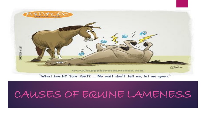
CAUSES OF EQUINE LAMENESS CAUSES OF EQUINE LAMENESS
TRAUMATIC INJURIES TRAUMATIC INJURIES SPRAINS, STRAINS, FRACTURES, MUSCULOSKELETAL INJURIES, NERVOUS SYSTEM DAMAGE
SPRAINS AND STRAINS SPRAINS AND STRAINS A sprain occurs when a sudden or severe twisting of a joint results in tearing or stretching of ligaments. A strain is usually less severe than a sprain and is the result of overstretching of ligaments or tendons through excessive use or improper movement. The most common damages are to the tendons and ligaments that run from the knee down to the foot including the superficial digital flexor tendon, the deep digital flexor tendon and the accessory and suspensory ligaments. The problem is most prevalent in hunters and jumpers, and horses with a swayed back are most commonly affected. Hind-quarter lameness and great sensitivity to pressure creating pain in the back are signs of overlapping or damaged spinous processes.
FRACTURES FRACTURES Fractures are another form of injury that are usually immediately recognizable, although in cases of some hairline and stress fractures especially those to the cannon bone and chip fractures involving the joints below the elbow and stifle or in the bones of the foot are not immediately obvious on visual inspection. Fractures are caused by accidents such as falls, being kicked by another horse, stepping into a hole. Horses are also subject to compression fractures or fractures caused by high torque forces on a limb. Bone fractures are usually classified as open or closed. An open fracture breaks through the skin and is readily observable. A closed fracture such as a simple fracture or a chip fracture is usually contained within the limb and becomes observable only when lameness, pain, swelling, or fever occurs. At one time, a diagnosis of a fracture meant either retirement or a death sentence for the horse. However, orthopedic techniques have advanced greatly and many fractures no longer carry a grim prognosis
MUSCULOSKELETAL INJURIES MUSCULOSKELETAL INJURIES Injuries to any part of the musculoskeletal system can result in lameness with tendon injuries being rather common in horses. Lacerated or ruptured tendons can occur in the legs and the feet usually from a deep cut, a fall, a kick from another horse, or damage caused by striking a stationary object. Tenosynovitis takes several forms depending on the location of the trauma and is distinguished by a sudden building of fluid within the sheath of the tendon accompanied by pain, heat and lameness. Septic tenosynovitis is the result of bacterial infection resulting in pus and inflammatory enzymes that can digest the tendon. Pain and lameness are severe.
MUSCULOSKELETAL INJURIES MUSCULOSKELETAL INJURIES Stringhalt involves the tendon of the lateral digital extensor muscle at the hock. It is characterized by a sudden upward jerking of the hind leg accompanied by an involuntary flexion of the hock as the horse steps forward. Some cases follow trauma involving the tendon, but in other cases, the cause is unknown. Bursitis is the result of trauma to a bursa which is a closed sac lined by a membrane that secretes a lubricating fluid. These sacs are located between moving parts of the limb and act as cushions to prevent friction. An acute bursitis causes lameness. Septic bursitis occurs when a bursa becomes infected with bacteria or sometimes fungi. Immediate treatment is necessary to prevent further complications. Bursitis affects shoulder joints, hips, the cunean tendon at the inside of the hock joint, the elbow, the knee and any other point of movement cushioned by a bursa, and is often evidenced by noticeable swelling in the area, as well as lameness.
DEGENERATIVE DISEASES DEGENERATIVE DISEASES ARTHRITIS/ DEGENERATIVE JOINT DISEASE/ OSTEOARTHRITIS
ARTHRITIS ARTHRITIS Arthritis, or what is commonly referred to as degenerative joint disease (DJD), takes a number of forms. It is the end result of various injurious processes. The disease usually begins with an inflamed joint capsule and can progress into erosion of the cartilage and fusion of the joint, Degenerative joint diseases affect both young and older horses. Stiffness and diminished range of motion are the hallmarks of lameness associated with arthritis. Acute serous arthritis is characterized by a swollen, tender, fluid-filled joint and is the result of either joint stress or injury. Also known as acute synovitis, this kind of arthritis does not necessarily progress to degenerative joint disease. Infectious or septic arthritis occurs when bacteria from the blood stream invade the joints destroying cartilage and causing irreversible damage.
ARTHRITIS ARTHRITIS Bone spavin is the name given to arthritis of the hock joint. It is an occupational hazard in horses that are ridden at a hard gallop such as jumpers, race horses, and hunters. Typically bone spavin disappears as a horse warms up and reappears when the horse cools down and is known as a " cold" lameness. Osselets is an arthritis of the fetlock joint and may affect one or both front feet. In the initial stages stretching or tearing of the fetlock joint capsule is accompanied by signs of acute serous arthritis. As the arthritis advances, pain and swelling occur along with new bone growth causing the horse to take short, choppy strides and plant weight on the outside edge of the hoof. Shoulder joint arthritis is another common form of arthritis. It often occurs after a fracture caused by being kicked by another horse, running into a stationary object, or by a hard fall. Osteochondrosis of the shoulder joint in growing horses may cause sufficient joint injury to lead to a degenerative arthritis.
FOOT RELATED LAMENESS FOOT RELATED LAMENESS FOOT WOUNDS, NAVICULAR DISEASE, POOR FOOT CONFORMATION, ROCKS, THRUSH, CANKER, QUITTOR, WHITE LINE DISEASE, HOOF WALL CRACKS
FOOT WOUNDS FOOT WOUNDS According to research most causes of lameness are found in the foot of the horse. Domesticated horses living in paddocks and stables with limited opportunity to toughen their feet are susceptible to a number of foot problems. Foot wounds that are contaminated and allow foreign bodies, bacteria, yeast, fungus, dirt, and debris to gain entrance to sensitive parts of the foot lead to abscesses and infections that cause lameness, especially if they are not noticed and treated promptly.
NAVICULAR DISEASE NAVICULAR DISEASE Navicular disease is noted as one of the leading causes of front leg lameness in horses. Foot stresses from hard stops, twists at high speeds, and abrupt changes in direction are thought to damage the navicular bone located at the heel of the foot beneath the frog.
POOR FOOT CONFORMATION POOR FOOT CONFORMATION Infrequent or inadequate hoof trimming results in a long toe and low heel, sheared heels, contracted heels and improper horse shoeing and are thought to adversely affect the transfer of weight through the navicular complex to the ground leading to injury to the inner structures of the foot. Initially, lameness is mild with navicular disease and comes and goes. As lameness worsens, a stiff, shuffling gait with a shortened, choppy stride becomes characteristic making it difficult for the horse to function.
ROCKS ROCKS Rocks and other hard objects can bruise the sole or lead to corns that will cause the horse to limp with lameness getting progressively worse if not treated promptly. In addition, if a sole bruise resulting in an abscess is allowed to become chronic, it can lead to pedal osteitis which is a thinning and demineralization of the coffin bone. Foot injuries can also result in keratomas which are tumors arising in the horn-producing cells of the hoof wall, usually in the toe region and sometimes in the sole. When the keratoma becomes large enough to cause lameness, it is usually surgically removed.
THRUSH THRUSH Thrush is a painful bacterial infection in the frog. Characterized by a putrid black discharge along with poor growth and degeneration of the horn, it is caused by lack of proper foot care resulting in a buildup of mud, manure and debris that prevents air getting to the frog.
CANKER CANKER Canker is a chronic infection of the horn tissues of the foot that begins at the frog and progresses into the sole and sometimes the hoof wall. It is caused by a lack of foot care, and is usually the result of the horse standing in mud or bedding that is soaked in urine and feces. The appearance is similar to thrush, but it involves the sole as well as the frog. Improper or neglected hoof trimming often contributes to lameness when a horse has or develops contracted heels or sheared heels. Usually corrective trimming and shoeing will prevent further lameness.
QUITTOR QUITTOR Quittor is another foot disease caused by a deep-seated infection of the cartilages of the coffin bone. Infected material is discharged via a sinus tract that opens at or above the coronet. Injuries such as being struck by another foot near the lateral cartilages and penetrating injuries of the sole lead to quittor.
WHITELINE DISEASE WHITELINE DISEASE Whiteline disease also known as seedy toe is caused when bacteria, yeast or fungus invades the foot and works its way up to the white line to the coronary band. Loss of horn creates a hollow space between the hoof wall and the sole that eventually is filled with cheesy material and debris. This disease seldom occurs in barefoot horses on pasture, but occurs with horses that are kept in wet stalls or exposed to frequent wet-to-dry episodes such as walking in wet grass or being washed down on a frequent basis.
HOOF WALL CRACKS HOOF WALL CRACKS Hoof wall cracks are another cause of lameness, although some hoof cracks do not cause the horse to go lame depending on the location and depth of the crack. Once a crack is noticed, care should be taken to prevent the crack from lengthening and deepening. Deep cracks are susceptible to infection and since new horn has to grow out from the coronary band to repair the crack, steps need to be taken to clean, stabilize, and either do corrective shoeing or repair the crack using prosthetic material.
DIET RELATED LAMENESS DIET RELATED LAMENESS LAMINITIS, AZOTURIA, NUTRITIONAL IMBALANCES
LAMINITIS LAMINITIS Laminitis, also known as founder, is a diet-related disease that commonly occurs when the horse consumes excess quantities of carbohydrates that alter the bacterial balance in the cecum, indirectly leading to the release of lactic acid and endotoxins. The lactic acid and endotoxins cause the large digital arteries to the feet to dilate, increasing the blood flow while causing intense constriction of the small capillaries that nourish the laminae in the horse's foot. Deprived of oxygen, the laminae swell, the swelling damages the sensitive tissue in the foot, and unless the situation is relieved the inner structure of the feet may die causing a characteristic stance in which the two front feet are placed out front to take weight off the horse's toes. The horse develops a high fever and chills with sweating, diarrhea, a fast pulse and rapid heavy breathing. The feet become hot and painful, and if all four feet are involved the horse may draw its feet up under its belly and lie down. Although death from founder is uncommon, it can occur. In cases of severe founder, the hoof may slough off.
LAMINITIS LAMINITIS Excess consumption of either too much grain or over-eating of lush, fast-growing summer pasture grasses causes most cases of founder. It can also occur during the winter if the horse consumes too much legume hay. Another cause of laminitis is the drinking of large amounts of cold water by an over-heated, hard working horse before being cooled down properly. Laminitis becomes chronic when lameness and pain continue for more than two days. Chronic laminitis can cause permanent damage to the foot when the coffin bone becomes detached from the hoof wall and rotates so that it drops down. In severe case it can penetrate the sole of the foot. Other complications of laminitis include white line disease, thrush, separations of the hoof at the coronary band or sole, and complete loss of the hoof. Acute laminitis is a medical emergency and a veterinarian should be called immediately to prevent the possibility of permanent lameness and disability.
AZOTURIA AZOTURIA Azoturia or tying-up syndrome are degrees of a condition known as exertional myopathy which occurs when horses that have a heavy workload have a break from activity, but continue to consume a high- carbohydrate diet. When an accumulation of glycogen builds up in the muscle as a result of lack of activity, lactic acid is released and damages skeletal muscle causing it to release muscle enzymes and myoglobin. When the myoglobin is excreted in the urine, it blocks the nephrons, causing kidney failure. The resultant lack of kidney function causes the horse to become anxious, sweat profusely and develop a rapid pulse. The major muscles then stiffen causing the horse to stagger and wobble and eventually to collapse. Upon the first indication of azoturia or tying-up, all activity should stop and the horse should be given absolute rest with no physical movement at all--not even returning to the stall. The horse should be spoken to calmly and covered with a blanket. A veterinarian should be called to provide medical relief to prevent further kidney damage and aid recovery.
NUTRITIONAL IMBALANCES NUTRITIONAL IMBALANCES Nutritional imbalances, especially in growing horses can lead to lameness caused by developmental orthopedic diseases which include a group of related conditions such as osteochondrosis, osteochondritis, dissecans, physeal dysplasia, and wobbler syndrome among others. In each case, a breakdown occurs in the mechanism by which cartilage is converted to bone. The resultant abnormal cartilage is prone to fracture, fissure and break into small fragments that can enter the joint causing lameness.
NUTRITIONAL IMBALANCES NUTRITIONAL IMBALANCES Although some horses appear to have a genetic predisposition for developing DOD, external factors include a too-high energy diet accompanied by insufficient calcium or phosphorus in the diet resulting in inadequate mineralization of bone. Also, a deficiency of microminerals such as copper and zinc, can result in defective cartilage formation. Generally, research shows that the most common feeding practices responsible for nutritional imbalances leading to lameness include: Feeding too much grain Feeding alfalfa hay without adding phosphorus to the ration Feeding a grass hay and grain mix inadequate in calcium, phosphorus, and protein. Adding excess vitamins and minerals to the ration
LIMB DEFORMITIES LIMB DEFORMITIES CONGENITAL LIMB DEFORMITIES, FLEXURAL LIMB DEFORMITIES
CONGENTIAL LIMB DEFORMITIES CONGENTIAL LIMB DEFORMITIES Congenital limb deformities are caused by abnormal limb positions in the uterus, nutritional imbalances in the mare, neonatal hypothyroidism or unequal growth between two sides of a long bone. Many normal foals have some degree of limb crookedness that straightens out by the time they become yearlings. Others need veterinarian treatment to correct the problem. Angular limb deformities include knock-knees, bow-legs, bucked or sprung knees, calf knees, benched or popped knees. Early recognition and treatment are important to prevent permanent damage. Treatment is usually based on x-rays and physical examination. Sometimes stall rest and therapeutic exercise are enough to correct the problem. Other cases may require splints, casts, braces or surgery. Treatment before three months of age works best in the prevention of future problems.
FLEXURAL LIMB DEFORMITIES FLEXURAL LIMB DEFORMITIES Flexural limb deformities tend to affect young horses from birth to 18 months of age. They occur most often in the fetlock joint, coffin joint and knee joint and involves a shortening of either the deep, the superficial, or both digital flexor tendons. The cause is unknown, but one theory is that rapid growth of long bones is not matched by the growth of the tendons, causing the joint to be pulled into flexion. Trauma or a developmental orthopedic disease such as osteochondrosis have also been implicated in flexural limb deformities, and in most cases evidence is found that nutritional excesses and imbalances are also involved.
FLEXURAL LIMB DEFORMITIES FLEXURAL LIMB DEFORMITIES In young foals, simply walking or exercising the foal several times a day will help stretch the tendon. Energy intake should be reduced and the ration carefully balanced. Trimming the heel helps to stretch and lengthen the tendon also. When the deformity is severe or progressing in spite of treatment, surgery is advised to divide the inferior check ligament thereby allowing the flexor tendon to lengthen. In some cases ligaments may be cut in advanced cases. In all cases, a veterinarian should be involved in diagnosing and treating the problem.