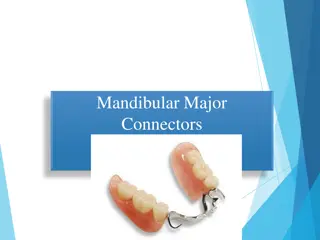
Mandibular Border Molding Techniques for Dental Impressions
Learn about the anatomical landmarks and sequence of border molding in mandibular denture fabrication, along with the importance of custom tray design and movements for accurate impressions. Explore the detailed steps involved in creating a well-fitted denture through passive and active movements in different flanges.

Download Presentation

Please find below an Image/Link to download the presentation.
The content on the website is provided AS IS for your information and personal use only. It may not be sold, licensed, or shared on other websites without obtaining consent from the author. If you encounter any issues during the download, it is possible that the publisher has removed the file from their server.
You are allowed to download the files provided on this website for personal or commercial use, subject to the condition that they are used lawfully. All files are the property of their respective owners.
The content on the website is provided AS IS for your information and personal use only. It may not be sold, licensed, or shared on other websites without obtaining consent from the author.
E N D
Presentation Transcript
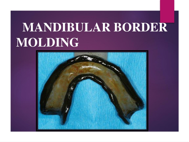
MANDIBULAR BORDER MOLDING
CUSTOM TRAY CUSTOM TRAY SHOULD BE 2 TO 3 MM SHORT OF VESTIBULE
SEQUENCE OF BORDER MOLDING LABIAL FLANGE 1. BUCCAL FLANGE 2. DISTOBUCCAL AREA AND MASETRIC NOTCHES 3. LINGUAL FLANGE 4. A] ANTERIORLINGUAL FLANGE B]MIDDLELINGUAL FLANGE C]DISTO LINGUAL FLANGE
TWO TYPES OF MOVEMENTS ACTIVE MOVEMENTS PASSIVE MOVEMENTS
LABIAL FLANGE PASSIVE MOVEMENTS LIP IS LIFTED OUTWORDS N INWORDS
BUCCAL FLANGE ACTIVE MOVENTS PATIENT IS ASKED TO PUCKER AND SMILE PASSIVE MOVEMENTS LIFT LIP OUTWARDS UPWARDS INWARDS
LINGUAL FLANGE ANTERIOR LINGUAL FLANGE MIDDLE LINGUAL FLANGE DISTOLINGUAL FLANGE
RETROMYLOHYOID CURTAIN Medial pterygoid and superior contrictor muscle is present in this space ACTIVATES SUPERIOR CONSTRICTOR PATIENT IS TOLD TO PROTRUDE THE TONGUE OUT CONTRACTION OF MEDIAL PETERYGOID MUSCLE ACTIVATES SUPERIOR CONSTRICTOR AND RECORDS POSTERIOR LATERALASPECT OF RETROMYLOHYOID CURTAIN PATIENT IS TOLD TO CLOSE UNDER PRESSURE
RETROMOLAR PAD Muscle involved 1. Posterior superior fibers of temporalis 2. Buccinator 3. Petrygomandibular Raphe 4. Medially by superior constrictor of pharynx ACTION OF ALL THE MUSCLES LEADS TO S SHAPED FLANGE AND PREVENTS OVER EXTENSION OF THE DENTURE BASE AND EVEN EXTRA IMPRESSION PRESSURE DURING