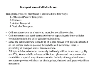
Membrane Fluorescent Tags and Fluidity
Membrane fluorescent tags can help study membrane fluidity by labeling lipids with fluorescent compounds. When exposed to UV light, tagged membranes glow uniformly unless treated with a laser beam, causing a bleached region. This activity reveals changes in membrane composition affecting fluorescence restoration time, indicating membrane fluidity alterations.

Download Presentation

Please find below an Image/Link to download the presentation.
The content on the website is provided AS IS for your information and personal use only. It may not be sold, licensed, or shared on other websites without obtaining consent from the author.If you encounter any issues during the download, it is possible that the publisher has removed the file from their server.
You are allowed to download the files provided on this website for personal or commercial use, subject to the condition that they are used lawfully. All files are the property of their respective owners.
The content on the website is provided AS IS for your information and personal use only. It may not be sold, licensed, or shared on other websites without obtaining consent from the author.
E N D
Presentation Transcript
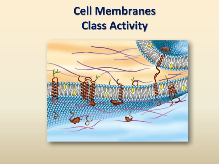
Cell Membranes Class Activity
Membrane Fluorescent tags and Fluidity Membrane lipids can be labelled with fluorescent tags by chemically attaching fluorescent compounds to phospholipids. When subjected to UV light, these fluorescent compounds absorb energy and then release it as light within the visible spectrum of the electromagnetic spectrum. When membranes that are tagged with fluorescent compounds are subjected to UV light, the entire surface of the cell will glow evenly. When shone on a tiny region of the cell, a strong focused laser light causes the denaturing of fluorescent tags and therefore loss of fluorescent activity in the subjected membrane region. Under UV light, this region looks dark giving the impression that there is a ``hole`` in the membrane or as if this region of the membrane has been ``bleached.``
B A
Figure 1. (A) Arrows indicate fluorescent cells whose membranes were treated with fluorescent tags. (D) A cell membrane surface. The dotted-line circle indicates an area that was subjected to a laser beam, before (-2s) and after the bleaching of the membrane (0s).
What do you think will happen to the bleached region when laser light is turned off? A. It will get bigger. B. It will get smaller, but will not disappear. C. It will get smaller and completely disappear. D. It will remain the same.
How can this be used to measure membrane fluidity?
Membrane Fluorescent tags and Fluidity This table shows data for cells with altered membrane composition. Cell membranes of the different cells were tagged with fluorescent compounds and then excited with UV light. The cells were then subjected to a localized focal laser beam in order to create a bleached region ( hole ) that can be examined under the microscope. The time it took the bleached region to become fluorescent again was recorded for each cell type. Time (s) for hole to become fluorescent 65 7 Condition No alteration length of fatty acid chains 38 2 cholesterol content unsaturation of fatty acid chains 88 9 42 2 membrane protein content 90 9
Which condition resulted in a membrane with highest fluidity? Time (s) for hole to become fluorescent 65 7 A. No alteration Condition B. length of fatty acid chains No alteration length of fatty acid chains C. cholesterol content 38 2 cholesterol content unsaturation of fatty acid chains D. unsaturation of fatty acid chains 88 9 42 2 E. membrane protein content membrane protein content 90 9
Using a graph, predict the relationship between cholesterol content and membrane fluidity (use Smart Board if available).
Youve just discovered an organism whose cell membranes are dominated by saturated phospholipids with extremely long hydrocarbon chains. In what type of habitat did you most likely find it? A. Salty B. Isotonic C. Hot D. Cold E. None of the above
Location of Membrane Proteins Lumen side Consider the epithelial cells lining the lumen of your small intestines. Nutrients and dietary minerals, such as glucose and Na+, are absorbed from the lumen side of the intestines, through the epithelial cells, and into blood vessels. For these substances to cross the membranes of epithelial cells, special membrane proteins (pumps, transporters, channels, etc.) are needed to be located strategically on the luminal and/or basal sides of the epithelial cells. Na+ Glucose Epithelial cell Basal side Blood flow
Location of Membrane Proteins Lumen side Predict the location of the following membrane proteins on the luminal and basal sides of intestinal epithelial cells and place them on the drawing of the cell shown above (use Smart Board). Justify your answers. Na/K pump Glucose/Na symporter GLUT- 1 transporter Na+ Glucose Epithelial cell Basal side Blood flow
Lumen side Na+ Glucose GLUT-1 Na+/glucose symporter Epithelial cell Basal side Na+/K+ pump Blood flow
Which of the following types of cell junctions is involved in determining the permanent location of membrane proteins in intestinal epithelial cells? A. Tight junction B. Desmosomes C. Gap junctions D. None of the above. Cell junctions do not determine the location of membrane proteins.


 Arrows indicate fluorescent cells whose")