Meta-analysis of Imaging Studies: Important Steps and Limitations
This meta-analysis delves into the significance of imaging studies, addressing the need for informed decisions, replication, and the challenges posed by underpowered analyses and prevailing experimental procedures. It explores the research question, inclusion/exclusion criteria, group effects, and the power of meta-analysis, providing a comprehensive overview aimed at guiding researchers in the field of neuroimaging.

Download Presentation

Please find below an Image/Link to download the presentation.
The content on the website is provided AS IS for your information and personal use only. It may not be sold, licensed, or shared on other websites without obtaining consent from the author.If you encounter any issues during the download, it is possible that the publisher has removed the file from their server.
You are allowed to download the files provided on this website for personal or commercial use, subject to the condition that they are used lawfully. All files are the property of their respective owners.
The content on the website is provided AS IS for your information and personal use only. It may not be sold, licensed, or shared on other websites without obtaining consent from the author.
E N D
Presentation Transcript
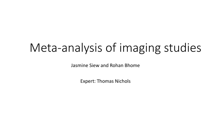
Meta-analysis of imaging studies Jasmine Siew and Rohan Bhome Expert: Thomas Nichols
Overview Why? Important steps Meta-analysis techniques Limitations/pitfalls
Background Single imaging studies underpowered analyses and thresholding procedures that increase false positives Influenced by experimental procedures So meta-analyses needed but: No clear consensus on neuroimaging meta-analysis Need for guidelines- informed and traceable decisions for researchers Replication
1. Research Question What is the research question? What approach to investigating it? Eg fMRI studies- do you include several tasks purporting to measure particular brain function or only one task to increase uniformity Decisions need to be made a priori Pre-register
Research question informs inclusion/exclusion criteria Inclusion Modalities of studies/ paradigms Subject groups- eg healthy controls only, studies with control groups, specific age range etc Analysis techniques- main effects vs interactions; papers reporting results on a particular threshold
Group Effects Between group effects: Experimental vs meta-analytical level Compare groups across all studies (eg Alzheimer s disease vs controls) or Separate meta-analyses for one group (Alzheimer s disease) vs other group (controls)
2. Power of the meta-analysis Presumption that enough studies have asked the research question for it to be worth doing a meta-analysis Small number of studies- one or two studies results may skew overall findings- limited generalisabilty Where large effect size expected- more acceptable to have fewer studies Trade off between number of studies and introducing heterogeneity Simulation study suggested atleast 17-20 experiments for smaller effect sizes
3. Data collection and extraction Literature search Combination of search terms: Disease ; Imaging modality ; experiments (if fMRI for eg) Reference lists of relevant articles Grey literature Search engines, search terms, date boundaries Data extraction sample size, x,y,z co-ordinates in standard space (i.e. MNI or TAL), whole-brain analysis, both increases and decreases in activation are reported (for SDM) Two investigators
4a. Included studies cover same search coverage CBMA Prerequisite. Usually whole brain. Generally avoid experiments with ROI or SVC analyses But this is a limitation as many studies with relevant findings may be ineligible. Could acknowledge in intro/discussion ROI Meta-analysis meta-analysis focusses on ROI in which case null-space has to be adapted to the ROI, i.e. testing against random spatial association across the ROI only All included experiments need to have used the same mask for ROI
4a. Included studies cover same search coverage (continued) Sometimes unclear if whole brain covered However, use scanner parameters to decide if whole brain covered or not Sometimes whole brain minus a few slices These experiments might be considered for inclusion, but should be reported as experiments with almost complete brain coverage
4b. Identify and adjust for differences in reference space Imaging data is normalized into a standard space in order to be able to investigate effects across subjects Talairach and Tournoux (TAL) and Montreal Neuroscience Institute(MNI) Different co-ordinates Identify from methods section of studies (sometimes unclear) Transform into same space
5. Adjust for multiple contrasts Multiple experiments for one set of subjects in a study Two ways: Adjust for within group differences- pool co-ordinates from all relevant contrasts and average contrast maps of sample and adjusting variance Combining contrast maps of sample using weighted mean depending on contribution of each contrast to each voxel
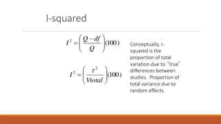
6. Find a balance between sensitivity and susceptibility to false positives controlling for the family-wise error (FWE) or the false discovery rate(FDR) on the voxel- or cluster-level ALE- cluster level FWE correction See later under meta-analysis techniques
Thomas Nichols, Neuroimaging Meta-Analysis OHBM Educational Course, 2018 Neuroimaging Meta-Analysis Approaches (1) Region of Interest Traditional Meta-Analysis, on mean %BOLD & stderr Almost impossible to do ROI-based results rare (exception: PET) Different ROIs used by different authors Heterogenous boundaries Peak %BOLD useless, due to voodoo bias Peak is overly-optimistic estimate of %BOLD in ROI True %BOLD Estimated %BOLD MNI x-axis
Intensity Based meta-analysis (IBMA) Access to statistical maps of whole brain data Subthreshold but consistent signal
Camille Maumet, Neuroimaging Meta-Analysis, OHBM Educational Course, 2017
IBMA (3)- statistical approaches With subjects statistics maps P/T/Z Images only Fisher s Stouffer s Stouffer s random effects- variability between studies Weighted Stouffer s- numbers in studies known; larger studies carry greater weight
IBMA (3) With Contrast of Parameter Estimate (COPE) only (Effect size images) Only allows random-effects model without weights With COPE s & VARCOPES (variance) Gold standard 3rd level mixed effects model FSL s FEAT/FLAME2nd-level FLAME: Combining subjects 3rd-level FLAME: Combining studies Allows meta-regression Units of Contrast maps need to be the same- different packages scale contrast data differently and this needs to be accounted for. Imaging data rarely shared and this is limiting.
Thomas Nichols, Neuroimaging Meta-Analysis OHBM Educational Course, 2018 Neuroimaging Meta-Analysis Approaches (3) Coordinate-Based Meta-Analysis (CBMA) x,y,z locations only Activation Likelihood Estimation (ALE) Turkeltaub et al. (2002). Meta-analysis of the functional neuroanatomy of single-word reading: method and validation. NeuroImage, 16(3), 765 780. Eickhoff et al. (2009). Coordinate-based activation likelihood estimation meta-analysis of neuroimaging data: a random-effects approach based on empirical estimates of spatial uncertainty. Human Brain Mapping, 30(9), 2907-26. Eickhoff et al. (2012). Activation likelihood estimation meta-analysis revisited. NeuroImage, 59(3), 2349 61 Multilevel Kernel Density Analysis (MKDA) Wager et al. (2004). Neuroimaging studies of shifting attention: a meta-analysis. NeuroImage 22 (4), 1679 1693. Kober et al. (2008). Functional grouping and cortical-subcortical interactions in emotion: a meta-analysis of neuroimaging studies. NeuroImage, 42(2), 998 1031. x,y,z and Z-value Signed Differential Mapping (SDM) Radua & Mataix-Cols (2009). Voxel-wise meta-analysis of grey matter changes in obsessive-compulsive disorder. British Journal of Psychiatry, 195:391-400. Costafreda et al. (2009). A parametric approach to voxel- based meta-analysis. NeuroImage, 46(1):115-122.
Thomas Nichols, Neuroimaging Meta-Analysis OHBM Educational Course, 2018 ALE ALE Activation Likelihood Estimation (Turkeltaub et al., 2002) ALE per-study map Study 1 Study 2 Study 3 Study 1 Study 2 Study 3 kernel FHWM f ALE map ALE interpretation for single focus Probability of observing a focus at that location ALE combining Probability of union of events ALE(p1,p2) = p1 + p2 p1 p2 ALE(p1,p2,p3) = p1 + p2 + p3 p1 p2 p1 p3 p2 p3 + p1 p2 p3
Eickoff et al., 2011 Compare observed ALE with null hypothesis ALE interpretation: Probability of observing one or more foci at a given location based on a model of Gaussian spread with FWHM (based on sample size) f
Multilevel Kernel Density Analysis Binary indicator maps for each contrast are created by convolving peak foci using a flat disc kernel Radius of kernel usually 10-15mm (Wagner et al., 2004) centred on focus Activation within distance r from location given a value of 1. MKDA statistic image is weighted based on number of participants in a study Robustness to possible bias from studies that systematically report more foci per cluster
MKDA (unweighted) interpretation: For any given location, what is proportion of studies having one or more foci within distance r Thomas Nichols, Neuroimaging Meta-Analysis OHBM Educational Course, 2018
Signed differential mapping (SDM) SDM is a stricter selection of the reported peak coordinates (only regions that appear statistically significant at the whole-brain level) Peak results that are corrected Co-ordinates converted from MNI to Talairach un-normalised Gaussian kernel around peak co-ordinates FWHM is set at 25mm Positive and negative activations/volumes recreated on same map - > therefore at meta-analytic level voxel will be insignificant
SDM (continued) null distribution created to test which voxels have more studies reporting differences around them than expected by chance. Whole brain level Monte Carlo randomisations Further iterations: anisotropic kernels (Radua et al., 2014) Permutation of subject images (PSI) (Radua et al., 2019).
Large scale meta-analysis tools Yarkoni, Poldrack, Nichols, Essen, & Wager (2011). Large-scale automated synthesis of human functional neuroimaging data. Nature Methods, 8(8), 665-670.
Use of large scale meta-analytic databases Use meta-analytic databases Where do foci occur for particular cognitive processes Then do the study
Reporting of meta-analysis Research question Detailed inclusion and exclusion criteria and the motivation why they were applied All steps of the meta-analytic study ideally in a flow-chart Number of experiments included in each analysis Handling of multiple experiments from the same subject group Detailed information on each included experiment (number of subjects, specific characteristics of the subjects, task description, stimuli used, coordinate space
Summary Muller et al., 2018
Pitfalls McInnes and Bussoyt, 2015


")

- statistical approaches")
")


 interpretation:")

")
")