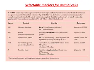
Metabolic Relationships in Cancer Cells
The figure illustrates the intricate connections between the metabolome, proteome, and genome in cancer cells. It highlights the dysregulated glycolysis, down-regulated TCA cycle, and pathways supporting cancer cell proliferation. Enzymes and oncogenes crucial for the cancer metabolic phenotype are also depicted. Understanding these metabolic changes is vital for targeted cancer therapies.

Download Presentation

Please find below an Image/Link to download the presentation.
The content on the website is provided AS IS for your information and personal use only. It may not be sold, licensed, or shared on other websites without obtaining consent from the author.If you encounter any issues during the download, it is possible that the publisher has removed the file from their server.
You are allowed to download the files provided on this website for personal or commercial use, subject to the condition that they are used lawfully. All files are the property of their respective owners.
The content on the website is provided AS IS for your information and personal use only. It may not be sold, licensed, or shared on other websites without obtaining consent from the author.
E N D
Presentation Transcript
WE Presentation #3, Lee et al Cell, A Lactate-Induced Response to Hypoxia 2015
Lee et al CELL 2015
The previous Figure illustrates the important relationships between metabolome, proteome, and genome in cancerous cells. Glycolysis breaks oxidizes glucose into 2 pyruvate, which is then fermented to lactate; pyruvate flux through the TCA cycle is down-regulated in cancer cells. Pathways branching off of glycolysis, such as the pentose phosphate pathway, generate biochemical building blocks to sustain the high proliferative rate of cancer cells. Blue boxes are enzymes important in transitioning to a cancer metabolic phenotype; orange boxes are enzymes that are mutated in cancer cells. Green ovals are oncogenes that are up-regulated in cancer; red ovals are tumor suppressors that are down-regulated in cancer. Figure abbreviations: 2PG: 2-phosphoglycerate; 3PG: 3-phosphoglycerate; BPG: 1,3-bisphosphoglycerate; CoA: coenzyme A; DHAP: dihydroxyacetone phosphate; F6P: fructose-6-phosphate; FBP: fructose-1,6-bisphosphate; G3P: glyceraldehyde-3- phospate; G6P: glucose-6-phosphate; HK: hexokinase; LDHA: lactate dehydrogenase A; PFK: phosphofructokinase; PI3K: phosphatidylinositide 3-kinase