Microbial Staining Techniques
Microbial staining techniques such as spore stain, capsule stain, and negative stain are essential in microbiology to visualize different structures of bacteria. Spore stains identify endospores formed by specific bacteria, while capsule stains highlight the protective capsules surrounding certain bacterial cells, aiding in pathogen identification.

Download Presentation

Please find below an Image/Link to download the presentation.
The content on the website is provided AS IS for your information and personal use only. It may not be sold, licensed, or shared on other websites without obtaining consent from the author.If you encounter any issues during the download, it is possible that the publisher has removed the file from their server.
You are allowed to download the files provided on this website for personal or commercial use, subject to the condition that they are used lawfully. All files are the property of their respective owners.
The content on the website is provided AS IS for your information and personal use only. It may not be sold, licensed, or shared on other websites without obtaining consent from the author.
E N D
Presentation Transcript
Spore stain Capsule stain
Endospores are formed by some bacteria, aerobic Bacillus species and anaerobic Clostridium species. Non active, resistant to heating and chemicals. Remain dormant until favorable conditions return.
Not easily stained Not easily stained: Mild heating. Use of strong stain. Timing of the stain is extended.
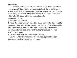
1-Primary stain: Malachite green, both vegetative cell and spores will appear green. 2-Decolorizing Agent: Water, it will remove the color from the cell, spores will remain green. 3-Counter stain: Safranin which will be absorbed by the vegetative cell.
Apply malachite green for 5 min application of heat is required. Rinse with D. water. Stain with safranin for 1 min. Observation: Spores appear green While vegetative bacteria or non spore forming bacteria appear red/pink
It is a negative stain where a dye does not have affinity for microbial cells but colors the background. Capsule is produced by some bacteria as a slippery substance that adheres to surface forming a viscous coat. This coat is called slime layer when it is loosely bound to bacterium.
Capsules are non ionic, uncharged because they are composed of polysaccharides This method use Nigrosin and Safranin to stain the bacteria and the background leaving capsule unstained as a clear halo surrounding bacteria. Capsule is important to some bacteria (pathogens).
Deliver 1 drop of Nigrosin by the loop and 1 drop of safranin at the end of clean slide. Mix a loopful of culture ( Kelbsiella sp.) with the stain. Spread the mixture on the slide as a blood film Air dry the film Leave the slide for 5 minutes at slide warmer to be fixed.