Microscopic Observation and Staining Techniques for Bacteria
Discover the process of microscopic observation and staining of microorganisms, focusing on size, shape, grouping, and staining reactions. Learn about the use of basic dyes to reveal internal and external structures of bacteria, as well as the preparation of fixed smears for study. Explore images depicting attraction between dyes and bacterial charges, bacterial smear preparation, simple staining, and gram staining to understand bacterial shapes.

Uploaded on | 1 Views
Download Presentation

Please find below an Image/Link to download the presentation.
The content on the website is provided AS IS for your information and personal use only. It may not be sold, licensed, or shared on other websites without obtaining consent from the author. If you encounter any issues during the download, it is possible that the publisher has removed the file from their server.
You are allowed to download the files provided on this website for personal or commercial use, subject to the condition that they are used lawfully. All files are the property of their respective owners.
The content on the website is provided AS IS for your information and personal use only. It may not be sold, licensed, or shared on other websites without obtaining consent from the author.
E N D
Presentation Transcript
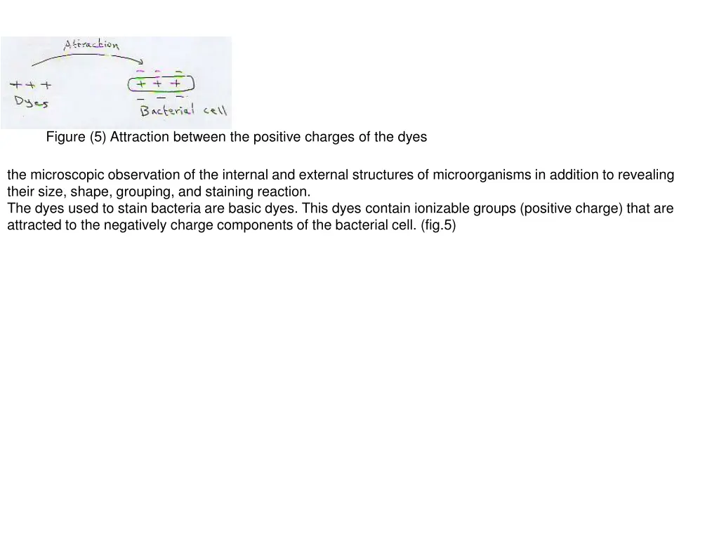
Figure (5) Attraction between the positive charges of the dyes the microscopic observation of the internal and external structures of microorganisms in addition to revealing their size, shape, grouping, and staining reaction. The dyes used to stain bacteria are basic dyes. This dyes contain ionizable groups (positive charge) that are attracted to the negatively charge components of the bacterial cell. (fig.5)
Preparation of fixed smears: (fig.6) 1- When the bacteria are taken from solid media, put one drop of water on clean side, suspend the bacteria with the drop of water and spread it over the slide. 2- Allow the smear to dry in air. 3- Fix the smear to the slide by passing rapidly through a flame for 2 to 3 times. Figure (6 Bacterial smear preparation
 Attraction between the positive charges of the")
")
 - Simple Staining and bacterial shapes.")