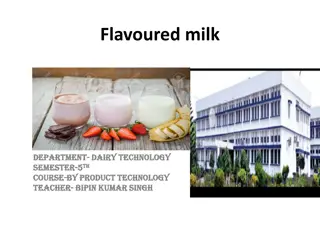
Milk Synthesis in Mammary Gland: Anatomy and Function
The mammary gland in cows plays a crucial role in milk synthesis, with specialized epithelial cells converting nutrients from blood into milk components. Understanding the anatomical structure and physiological processes involved in milk production is essential for maintaining quality and preventing issues like mastitis. Explore how the mammary gland functions, from synthesis to secretion, and the intricate mechanisms behind milk biosynthesis.

Download Presentation

Please find below an Image/Link to download the presentation.
The content on the website is provided AS IS for your information and personal use only. It may not be sold, licensed, or shared on other websites without obtaining consent from the author.If you encounter any issues during the download, it is possible that the publisher has removed the file from their server.
You are allowed to download the files provided on this website for personal or commercial use, subject to the condition that they are used lawfully. All files are the property of their respective owners.
The content on the website is provided AS IS for your information and personal use only. It may not be sold, licensed, or shared on other websites without obtaining consent from the author.
E N D
Presentation Transcript
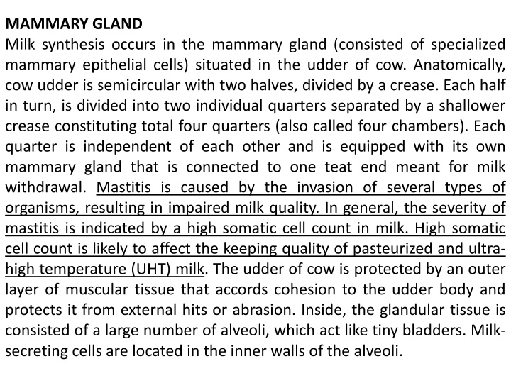
MAMMARY GLAND Milk synthesis occurs in the mammary gland (consisted of specialized mammary epithelial cells) situated in the udder of cow. Anatomically, cow udder is semicircular with two halves, divided by a crease. Each half in turn, is divided into two individual quarters separated by a shallower crease constituting total four quarters (also called four chambers). Each quarter is independent of each other and is equipped with its own mammary gland that is connected to one teat end meant for milk withdrawal. Mastitis is caused by the invasion of several types of organisms, resulting in impaired milk quality. In general, the severity of mastitis is indicated by a high somatic cell count in milk. High somatic cell count is likely to affect the keeping quality of pasteurized and ultra- high temperature (UHT) milk. The udder of cow is protected by an outer layer of muscular tissue that accords cohesion to the udder body and protects it from external hits or abrasion. Inside, the glandular tissue is consisted of a large number of alveoli, which act like tiny bladders. Milk- secreting cells are located in the inner walls of the alveoli.
Secreted milk is carried by capillaries into milk ducts, which lead into cavity known as cistern, located above the teat. Cistern holding about one third of milk of the udder, is connected to teat. The teat contains a channel that allows milk to exit from teat. A sphincter muscle is located at end of the teat channel to close the flow of milk between milking. The muscle prevents milk from leaking out of the teat and controls entry of bacteria and other harmful microbes. During hand or machine milking, the hormone oxytocin allows let down of milk and initiates emptying of milk from udder. Preparation of cow for milking includes stimulation of udder by calf suckling or by other stimuli in the milking parlor signals release of the hormone into blood. The let down of milk by oxytocin is complete in less than a minute. The muscle-like cells of alveoli develop pressure in the udder, forcing milk into teat cistern from where it can be withdrawn by suction of milking machine or by applying pressure on the teat during hand milking. The effect of oxytocin lasts for 5 8 minutes, during which milking process should be completed.
Mammary gland has a sophisticated level of organization with a remarkable ability to convert circulating nutrients from blood into milk components. Mammary epithelial cells in mammary gland synthesize complex milk constituents from simple components present in circulating blood. Mammary gland is equipped with an extensive network of arterial and venous blood capillaries. The components of blood needed for milk biosynthesis are extracted from arterial blood. The venous blood carries the blood back to the heart for recirculation and component renewal. Precursors of milk proteins, lipids, and carbohydrates are present in blood. All the milk fat, lactose, and most milk proteins are then synthesized by the mammary epithelial cells. Certain milk constituents (milk fat, most milk proteins) are specific to specie of the animal. Lactose is also synthesized in the mammary gland but it is identical in all species of mammals. Water, salts, vitamins, immunoglobulins, some hormones, and serum albumin leak into milk unchanged from blood. It is generally agreed that to synthesize 1 liter of milk, the epithelial cells of mammary gland require 800 900 liters of blood.
moving through them for providing adequate level of milk precursors. Lactating mammary gland extracts glucose, amino acids, fatty acids, - hydroxy butyrate, and mineral salts from blood for synthesis of milk constituents. The epithelial cells synthesize milk continuously from the precursors and store milk until it is removed. The ratios of major milk and blood components suggest that fat, sugar, potassium, calcium, magnesium, and phosphate are several times more abundant in milk than in blood. However, the concentration of protein, phospholipid, sodium, and chloride is significantly more in blood than in milk. BIOSYNTHESIS OF MILK PROTEINS For biosynthesis of milk constituents, the primary substrates extracted by mammary epithelial cells from their counterparts in blood include glucose, amino acids, fatty acids, -hydroxy butyrate, and salts. Knowledge relative to biosynthesis of milk constituents is not fully understood; therefore, it is an evolving science. In the ruminant animals, all food must pass through rumen prior to digestion in the stomach and intestines.
A large proportion of dietary protein is transformed by rumen bacteria and protozoa, thereby generating high-quality microbial protein with significantly better amino acid profile than that of the vegetable protein in the feed. After digestion, the microbial protein along with smaller quantity of feed protein (that escaped rumen digestion) gives rise to small peptides and amino acids. These are then transported across the intestinal wall into blood, which ultimatel. The biosynthesis of milk proteins in mammary gland resembles biosynthesis of any other protein in body tissues. The substrate, amino acids from blood, is transported through the basolateral membrane to mammary secretory cell.
BIOSYNTHESIS OF MILK LIPIDS Milk fat in freshly secreted milk occurs as microscopic globular emulsion of liquid fat in aqueous phase of milk plasma. Fat content of milk varies from 3.4 to 5.1%, depending on the breed of the cow. Most of the milk used in dairy processing typically contains an average of 3.5 3.6% fat. Variability of milk fat depends upon the individuality of animal, stage of lactation, feed, environmental factors, and stage of milking. The functional properties of milk fat are attributed to its fatty acid make up. Milk fat functions as a concentrated source of energy as well as a source of fat-soluble vitamins A, D, E, and K and essential fatty acids, linoleic, and arachidonic acids. The essential fatty acids are not synthesized by human body. They must be supplied by the diet. Arachidonic acid with four double bonds is present in traces. Its precursor is linoleic acid. The biosynthesis of individual fatty concentration in milk fat has a profound effect on properties and utilization of milk fat. Saturated fatty acids are solid at ambient temperature, while unsaturated fatty acids are liquid. acids and their comparative
Their ratio in milk fat has a significant effect on the hardness and spreadability of butter at refrigerated Furthermore, the balance between C4 and C18 fatty acids keeps milk fat liquid at body temperature. BIOSYNTHESIS OF MILK SUGAR, LACTOSE Glucose is the exclusive monosaccharide substrate for lactose biosynthesis. In ruminants, 45 60% of blood glucose is formed from propionate in the liver by Gluconeogenesis process. It is interesting that blood glucose level in nonruminants is approximately double than in the blood of ruminants. Biosynthesis of lactose occurs in the membranes of Golgi apparatus. Two molecules of glucose give rise to one molecule of lactose. Glucose is converted to UDP galactose by a cascade of several enzymatic reactions. At the onset of parturition, the enzyme activity shows a dramatic increase to cope up with lactogenesis (the lactation process). Glucose and UDP-galactose are combined to form lactose, catalyzed by the action of lactose synthase that is composed of galactosyl transferase and -lactalbumin. storage temperature.
SECRETION OF MILK CONSTITUENTS INTO LUMEN Milk constituents are individually synthesized inside the secretory cell. After they are transported to lumen space, they blend together to form so-called milk. The process for secreting nonfat constituents differs from that of milk lipids. Milk proteins synthesized in the RER (Rough Endoplasmic Reticulum )are incorporated into Golgi vacuoles or vesicles along with lactose and minerals. The secretory vesicles then separate from the Golgi apparatus and transport molecules toward the apical region of the cell. The membrane surrounding the vesicles fuses with the plasma membrane of epithelial cells followed by delivery into lumen space. At this point, major minerals are partitioned into colloidal and solution phase. Calcium and phosphate are associated with casein micelles. Milk lipids follow a discrete secretary process. As the molecules of synthesized milk fat transfer from the endoplasmic reticulum toward the apical membrane, their droplets grow in size. While passing through the apical membrane, they are pinched off as spherical globules with a coating of apical plasma membrane.
Bioactive Peptides Since time immemorial, milk and milk products have provided nutritional and energy needs for the milk consuming consumer. However, there is now increasing realization that diet could influence health. As a result, health-promoting benefits are sought by modern consumer in their food menu. In this context, bioactive properties displayed by some proteins, peptides, and fatty acids in milk has attracted strong research interest. Among milk components, CLA, vaccenic acid, and sphingolipids are thought to be associated with anticancer properties. Also, CLA, stearic acid, and omega-3 fatty acids are associated with improved cardiovascular health. Antihypertensive peptides ( -s1-casokinin, -casokinin, and -lactorphin) in milk are associated with cardiovascular health through inhibition of angiotensin converting enzyme. CLA along with calcium, phosphorus, and probiotics tends to improve bone health and immune protection. It is reasonable to conjecture that new era of functional foods and health awareness would continue to provide impetus to drive research in this fascinating biopharma interface of dairy industry.
Fighting Intramammary Infections Since udder health is important for uniform compositional quality of milk, efforts are ongoing to enhance mastitis control and prevention strategies. Good on farm mastitis control measures have addressed contagious mastitis problem; however, attributed to Streptococcus uberis and Escherichia coli remain a challenge. Immunotherapeutic strategy employing efficacious and specific protective antibody in mammary mucosa holds considerable Promise. Researchers at the University of Vermont have enabled mammary gland cells to secret antibacterial factor lysostaphin, which is antibacterial for Staphylococcus aureus. environmental mastitis