Learn about mitral stenosis, a condition that obstructs blood flow in the heart due to thickening of the mitral valve leaflets. Discover the pathophysiology, clinical manifestations, and effects on cardiac function. Find out about the University of Basrah College of Nursing's insights on this condition.
Please find below an Image/Link to download the presentation.
The content on the website is provided AS IS for your information and personal use only. It may not be sold, licensed, or shared on other websites without obtaining consent from the author. If you encounter any issues during the download, it is possible that the publisher has removed the file from their server.
You are allowed to download the files provided on this website for personal or commercial use, subject to the condition that they are used lawfully. All files are the property of their respective owners.
The content on the website is provided AS IS for your information and personal use only. It may not be sold, licensed, or shared on other websites without obtaining consent from the author.
E N D
Presentation Transcript
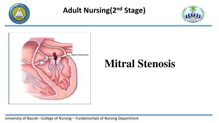
Adult Nursing(2nd Stage) Mitral Stenosis University of Basrah College of Nursing Fundamentals of Nursing Department
Mitral Stenosis Is an obstruction to blood flowing from the left atrium into the left ventricle . It most often is caused by rheumatic endocarditis, which progressively thickens mitral valve leaflets. Leaflets often fuse together. Eventually, the mitral valve orifice narrows and progressively obstructs blood flow into the ventricle. University of Basrah College of Nursing Fundamentals of Nursing Department
Pathophysiology Normally, the mitral valve orifice is as wide as the diameter of three fingers. In cases of severe stenosis, the orifice narrows to the width of a pencil. The left atrium has difficulty moving blood into the ventricle because of increased resistance by the narrowed orifice. Poor left ventricular filling can cause decreased cardiac output. Increased blood volume in the left atrium causes it to dilate and hypertrophy. Because there is no valve to protect pulmonary veins from backward flow of blood from the atrium, the pulmonary circulation becomes congested. As a result, the right ventricle must contract against abnormally high pulmonary arterial pressure and is subjected to excessive strain. University of Basrah College of Nursing Fundamentals of Nursing Department
Pathophysiology The right ventricle hypertrophies, eventually dilates, and fails. If the heart rate increases, diastole is shortened; thus, the amount of time for forward flow of blood is less, and more blood backs into the pulmonary veins. Therefore, as the heart rate increases, cardiac output decreases and pulmonary pressures increase. University of Basrah College of Nursing Fundamentals of Nursing Department
Clinical Manifestations 1-The first symptom of mitral stenosis often is dyspnea on exertion (DOE) as a result of pulmonary venous hypertension. 2-Patients may experience progressive fatigue and decreased exercise tolerance as a result of low cardiac output. 3-An enlarged left atrium may create pressure on the left bronchial tree, resulting in a dry cough or wheezing. 4-Patients may expectorate blood (i.e., hemoptysis) or experience palpitations, orthopnea, paroxysmal nocturnal dyspnea (PND), and repeated respiratory infections. 5-As a result of increased blood volume and pressure, the atrium dilates, hypertrophies, and becomes electrically unstable (patients experience atrial dysrhythmias). University of Basrah College of Nursing Fundamentals of Nursing Department
Causes Rheumatic fever: Rheumatic fever is the most common cause of mitral valve stenosis. It can damage the mitral valve by causing the flaps to thicken or fuse. Calcium deposits: calcium deposits can build up around the mitral valve, which can occasionally cause mitral valve stenosis. Radiation therapy. Treatment for certain types of cancer that requires radiation to chest area can sometimes cause the mitral valve to thicken and harden. Other causes. In rare cases, babies are born with a narrowed mitral valve (congenital defect) that causes problems over time. Some autoimmune diseases, such as lupus, also may rarely cause mitral valve stenosis. University of Basrah College of Nursing Fundamentals of Nursing Department
Assessment and Diagnostic Findings 1-The pulse is weak and irregular in the presence of atrial fibrillation (caused by strain on the atrium). 2-A low-pitched, rumbling diastolic murmur is heard at the apex and the patient may have signs or symptoms of heart failure. 3-Echocardiography is used to diagnose and quantify the severity of mitral stenosis. 4-Electrocardiography (ECG), exercise testing, and cardiac catheterization with angiography may be used to help determine the severity of mitral stenosis. University of Basrah College of Nursing Fundamentals of Nursing Department
Medical Management 1-Patients with mitral stenosis may benefit from anticoagulants to decrease the risk of developing atrial thrombus and may require treatment for angina. 2-If atrial fibrillation develops, cardioversion is attempted to restore normal sinus rhythm. If unsuccessful, the ventricular rate is controlled with beta blockers, digoxin, or calcium channel blockers. 3-Surgical intervention consists of valvuloplasty, usually a commissurotomy to open or rupture the fused commissures of the valve. 4- Percutaneous transluminal valvuloplasty or valve replacement may be performed. University of Basrah College of Nursing Fundamentals of Nursing Department
Complications High blood pressure in the lung arteries (pulmonary hypertension). Increased pressure in the arteries that carry blood from heart to lungs (pulmonary arteries) causes heart to work harder. Heart failure. A narrowed mitral valve interferes with blood flow. As a result, pressure may increase in lungs, leading to fluid buildup. The fluid buildup strains the right side of the heart, leading to right heart failure. Fluid in the lungs (pulmonary edema). Heart enlargement. The pressure buildup of mitral valve stenosis results in enlargement of heart's upper left chamber (atrium). University of Basrah College of Nursing Fundamentals of Nursing Department
Complications Atrial fibrillation. The stretching and enlargement of left atrium may lead to rhythm problem in which the upper chambers of heart beat too quickly. Blood clots. Untreated atrial fibrillation can cause blood clots to form in the upper left chamber of heart. Blood clots from heart can break loose and travel to other parts of body, causing serious problems, such as a stroke if a clot blocks a blood vessel in brain. University of Basrah College of Nursing Fundamentals of Nursing Department
Aortic Regurgitation Is flow of blood back into the left ventricle from the aorta during diastole. It may be caused by inflammatory lesions that deform aortic valve leaflets or dilation of the aorta, preventing complete closure of the aortic valve. This valvular defect also may result from infective or rheumatic endocarditis, congenital abnormalities, diseases such as syphilis, blunt chest trauma, or deterioration of a surgically replaced aortic valve. University of Basrah College of Nursing Fundamentals of Nursing Department
Pathophysiology Blood from the aorta returns to the left ventricle during diastole, in addition to blood normally delivered by the left atrium. The left ventricle dilates in an attempt to accommodate the increased volume of blood. It also hypertrophies in an attempt to increase muscle strength to expel more blood with above-normal force, thus increasing systolic blood pressure. Arteries attempt to compensate for higher pressures by reflex vasodilation; peripheral arterioles relax, reducing peripheral resistance and diastolic blood pressure. University of Basrah College of Nursing Fundamentals of Nursing Department
Clinical Manifestations 1-Some patients are aware of a forceful heartbeat, especially in the head or neck. 2-Marked arterial pulsations visible or palpable at carotid or temporal arteries may be present as a result of increased force and volume of blood ejected from a hypertrophied left ventricle. 3-Exertional dyspnea and fatigue follow. Signs and symptoms of progressive left ventricular failure include breathing difficulties (e.g., orthopnea, PND). University of Basrah College of Nursing Fundamentals of Nursing Department
Assessment and Diagnostic Findings 1-A high-pitched, blowing diastolic murmur is heard at the third or fourth intercostal space at the left sternal border. 2-The pulse pressure (i.e., difference between systolic and diastolic pressures) is considerably widened in patients with aortic regurgitation. 3-One characteristic sign is the water hammer (Corrigan s) pulse, in which the pulse strikes a palpating finger with a quick, sharp stroke and then suddenly collapses. 4-The diagnosis may be confirmed by echocardiography, cardiac magnetic resonance imaging (MRI), and cardiac catheterization 5-Patients with symptoms usually have echocardiograms every 6 months, and those without symptoms have echocardiograms every 2 to 5 years. University of Basrah College of Nursing Fundamentals of Nursing Department
Nursing Management 1-The patient is instructed to report new symptoms or changes in symptoms to the primary provider. 2-Measures the patient s heart rate, blood pressure, and respiratory rate, compares these results with previous data, and notes any changes. 3-Heart and lung sounds are auscultated and peripheral pulses palpated. 4-The nurse assesses the patient for the following: Signs and symptoms of heart failure, such as fatigue, DOE, decreased activity tolerance, an increase in coughing, hemoptysis, multiple respiratory infections, orthopnea, and PND. Dysrhythmias, by palpating the patient s pulse for strength and rhythm (i.e., regular or irregular) and asking whether the patient has experienced palpitations or felt forceful heartbeats. -Symptoms such as dizziness, syncope, increased weakness, or angina pectoris University of Basrah College of Nursing Fundamentals of Nursing Department
5- Collaborates with the patient to develop a medication schedule and provides education about the name, dosage, actions, adverse effects, and any drug drug or drug food interactions of prescribed medications for heart failure, dysrhythmias, angina pectoris, or other symptoms. 6-Instructs the patient about the importance of attempting to relieve the symptoms of angina with rest and relaxation before taking nitroglycerin and to anticipate the potential adverse effects. 7-Assist the patient with planning activity and rest periods to achieve an acceptable lifestyle. Patients who experience symptoms of pulmonary congestion are advised to rest and sleep sitting in a chair or bed with the head elevated. 8- Educates the patient to take a daily weight and report sudden weight gain. University of Basrah College of Nursing Fundamentals of Nursing Department


")


