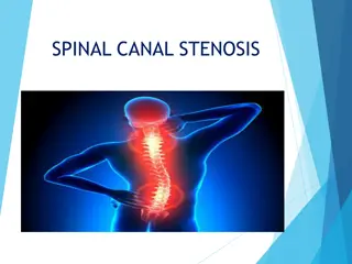
Mitral Stenosis Pathophysiology and Complications
Mitral stenosis is primarily caused by rheumatic disease, leading to fibrosis, calcification, and fusion of the mitral valve apparatus. This results in increased left atrial pressure, pulmonary congestion, and breathlessness. Progressive dilatation of the left atrium can trigger atrial fibrillation, exacerbating hemodynamic instability and pulmonary edema. Pulmonary hypertension may develop, providing some protection against pulmonary edema but leading to right ventricular hypertrophy.

Download Presentation

Please find below an Image/Link to download the presentation.
The content on the website is provided AS IS for your information and personal use only. It may not be sold, licensed, or shared on other websites without obtaining consent from the author.If you encounter any issues during the download, it is possible that the publisher has removed the file from their server.
You are allowed to download the files provided on this website for personal or commercial use, subject to the condition that they are used lawfully. All files are the property of their respective owners.
The content on the website is provided AS IS for your information and personal use only. It may not be sold, licensed, or shared on other websites without obtaining consent from the author.
E N D
Presentation Transcript
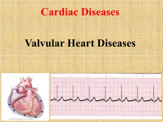
Valvular Heart Disease Dr. Zaki Bettamer Zikoecho@yahoo.com 3/13/2025 2:31 PM
73 years old man complains of chest pain and dyspnea while climbing stairs . On examination : BP: 120/90 mmhg, pulse : 75 beat / minute, slow rising carotid pulse which diminished on volume with Ejection systolic murmur radiates to the neck. The best diagnostic tool in this patient is
Mitral valve disease Mitral stenosis Aetiology almost always rheumatic in origin. heavy calcification of the mitral valve apparatus ( Old people ) Congenital (rare)
pathophysiology mitral stenosis MV fibrosis, calcification , and fusion and subvalvular apparatus. flow of blood from LA to LV and left atrial pressure rises. pulmonary venous congestion and breathlessness. dilatation and hypertrophy of the LA, and left ventricular filling becomes more dependent on left atrial contraction
Any in heart rate shortens diastole when the mitral valve is open and produces a further in left atrial pressure. Situations that demand in cardiac output also left atrial pressure, so exercise and pregnancy are poorly tolerated
The mitral valve orifice is normally about 5 cm 2 in diastole and may be reduced to 1 cm 2 in severe mitral stenosis. Patients usually remain asymptomatic until the stenosis is less than 2 cm 2 . Reduced lung compliance, due to chronic pulmonary venous congestion, contributes to breathlessness, and a low cardiac output may cause fatigue.
Atrial fibrillation due to progressive dilatation of the LA is very common. Its onset often precipitates pulmonary oedema because the accompanying tachy cardia and loss of atrial contraction lead to marked haemodynamic deterioration with a rapid rise in left atrial pressure.
In contrast, a more gradual in left atrial pressure tends to cause an in pulmonary vascular resistance, which leads to pulmonary hypertension that may protect the patient from pulmonary oedema. Pulmonary hypertension leads to right ventricular hypertrophy and dilatation, tricuspid regurgitation and right heart failure.
Clinical features Symptoms Breathlessness (pulmonary congestion) Fatigue (low cardiac output) Oedema, ascites (right heart failure) Palpitation (atrial fibrillation) Haemoptysis (pulmonary congestion, pulmonary embolism) Cough (pulmonary congestion) Chest pain (pulmonary hypertension) Thromboembolic complications (e.g. stroke, ischaemic limb)
Signs Atrial fibrillation Mitral facies Auscultation Loud first heart sound, opening snap Mid-diastolic murmur Crepitations, pulmonary oedema, effusions (raised pulmonary capillary pressure) RV heave, loud P 2 (pulmonary hypertension)
Investigations ECG Right ventricular hypertrophy: tall R waves in V 1 V 3 P mitrale or atrial fibrillation Chest X-ray Enlarged LA and appendage Signs of pulmonary venous congestion
Echo Thickened immobile cusps Reduced valve area Enlarged LA Reduced rate of diastolic filling of LV Doppler Pressure gradient across mitral valve Pulmonary artery pressure Left ventricular function Cardiac catheterisation Coronary artery disease Pulmonary artery pressure Mitral stenosis and regurgitation
Distended pulmonary veins Enlarged pulmonary trunk Splaying of carina Enlarged left atrium Double shadow of left atrial enlargement1 `
Management minor symptoms treated medically. symptomatic despite medical treatment or pulmonary hypertension balloon valvuloplasty, mitral valvotomy or mitral valve replacement should be considered .
Medical management anticoagulation to reduce the risk of systemic embolism. ventricular rate control (digoxin, -blockers or rate limiting calcium antagonists) in atrial fibrillation. diuretic to control pulmonary congestion. Antibiotic prophylaxis against infective endocarditis is no longer routinely recommended.
Mitral balloon valvuloplasty & valve replacement Criteria for mitral valvuloplasty. Significant symptoms. Isolated mitral stenosis. No mitral regurgitation. Mobile, non-calcified valve on echo. LA free of thrombus.
mitral valvuloplasty or valvotomy should be followed up at 1 2-yearly intervals because restenosis may occur. Clinical symptoms and signs are a guide to the severity of mitral restenosis but Doppler echocardiography provides a more accurate assessment.
Valve replacement is indicated if there is substantial mitral reflux or if the valve is rigid and calcified .
Mitral regurgitation Causes of mitral regurgitation Mitral valve prolapse Dilatation of the LV and mitral valve ring (e.g. coronary artery disease, cardiomyopathy) Damage to valve cusps and chordae (e.g. rheumatic heart disease, endocarditis) Ischemia or infarction of the papillary muscle Myocardial infarction
pathophysiology Chronic MR causes gradual dilatation of the LA with little increase in pressure and therefore relatively few symptoms. LV dilates slowly and the left ventricular diastolic and left atrial pressures gradually increase as a result of chronic volume overload of the LV. In acute MR causes a rapid rise in left atrial pressure and marked symptomatic deterioration.
Mitral valve prolapse (floppy ) common causes of mild MR. congenital anomalies or degenerative changes. sometimes a feature of connective tissue disorders such as Marfan s syndrome . mildest forms, the valve remains competent but bulges back into the atrium during systole, causing a mid-systolic click but no murmur.
M V P presence of aregurgitant valve, the click is followed by a late systolic murmur, which lengthens as the regurgitation becomes more severe. MVP is associated with a variety of typically benign arrhythmias, atypical chest pain and very small risk of embolic stroke or TIA. long-term prognosis is good.
Clinical features Symptoms Dyspnoea (pulmonary venous congestion) Fatigue (low cardiac output) Palpitation (atrial fibrillation, increased stroke volume) Oedema, ascites (right heart failure)
Signs Atrial fibrillation/flutter Cardiomegaly: displaced hyperdynamic apex beat Apical pansystolic murmur thrill Soft S1, apical S3 Signs of pulmonary venous congestion (crepitations,pulmonary oedema, effusions) Signs of pulmonary hypertension and right heart failure
Investigations ECG Left atrial hypertrophy (if not in atrial fibrillation) Left ventricular hypertrophy AF Chest X-ray Enlarged LA Enlarged LV Pulmonary venous congestion Pulmonary oedema (if acute)
Echo Dilated LA, LV Dynamic LV Structural abnormalities of mitral valve (e.g. prolapse) Doppler Detects and quantifies regurgitation Cardiac catheterisation Dilated LA, dilated LV, mitral regurgitation Pulmonary hypertension Coexisting coronary artery disease
Management Mitral regurgitation of moderate severity can be treated medically Diuretics Vasodilators ( after loud, HTN ) e.g. ACE inhibitors Digoxin if atrial fibrillation Anticoagulants if atrial fibrillation
Patients should be reviewed at regular intervals worsening symptoms progressive cardiomegaly or echocardiographic evidence of deteriorating left ventricular function mitral valve replacement or repair.
Aortic valve disease Aortic stenosis Aetiology Infants, children, adolescents Congenital AS Congenital subvalvular AS Congenital supravalvular AS Young adults to middle aged Calcification&fibrosis of congenitally bicuspid AV. Rheumatic AS.
Middle-aged to elderly Senile degenerative aortic stenosis Calcification of bicuspid valve Rheumatic AS.
pathophysiology COP is initially maintained at the cost of asteadily increasing pressure gradient across the AV. The LV becomes increasingly hypertrophied and coronary blood flow may then be patients may therefore develop angina, even in the absence of concomitant coronary disease.
The fixed outflow obstruction limits the increase in cardiac output required on exercise. the LV can no longer overcome the outflow tract obstruction and pulmonary oedema supervenes.
Clinical features Symptoms Mild or moderate stenosis: usually asymptomatic Exertional dyspnoea Angina Exertional syncope Sudden death Episodes of acute pulmonary oedema
Signs Ejection systolic murmur Slow-rising carotid pulse Thrusting apex beat (LV pressure overload) Narrow pulse pressure Signs of pulmonary venouscongestion (e.g. crepitations)
Investigations ECG Left ventricular hypertrophy (usually) Left bundle branch block Chest X-ray May be normal; sometimes enlarged LV and dilated ascending aorta on PA view, calcified valve on lateral view Echo Calcified valve with restricted opening, hypertrophied LV
Doppler Measurement of severity of stenosis Detection of associated aortic regurgitation Cardiac catheterisation Mainly to identify associated coronary artery disease May be used to measure gradient between LV and aorta
Management Irrespective of the severity of valve stenosis, patients with asymptomatic AS have a good immediate prognosis and conservative management . review, as the development of angina, syncope, symptoms of low cardiac output or heart failure has a poor prognosis and is an indication for surgery.
moderate or severe stenosis are evaluated every 1 2 years with Doppler echocardiography to detect progression in severity; this is more rapid in older patients with heavily calcified valves. symptomatic severe AS should have prompt aortic valve replacement. Aortic balloon valvuloplasty is useful in congenital AS ( but not in older patients with calcific AS ). Anticoagulants ( AF or AVR )
Aortic stenosis in old age Incidence: the most common form of valve disease affecting the very old. Symptoms: a common cause of syncope, angina and heart failure in the very old. Signs: because of increasing stiffening in the central arteries, low pulse pressure and a slow rising pulse may not be present.
Surgery: can be successful in those aged 80 yrs or more in the absence of comorbidity, but with a higher operative mortality. The prognosis without surgery is poor once symptoms have developed. Valve replacement type: a biological valve is often preferable to a mechanical one because this obviates the need for anticoagulation, and the durability of biological valves usually exceeds the patient s anticipated life expectancy.
Aortic regurgitation Causes of aortic regurgitation Congenital Bicuspid valve or disproportionate cusps Acquired Rheumatic disease Infective endocarditis Trauma Aortic dilatation (Marfan s syndrome, aneurysm, dissection, syphilis, ankylosing spondylitis)
pathophysiology This condition is due to disease of the aortic valve cusps or dilatation of the aortic root . The LV dilates and hypertrophies to compensate for the regurgitation. The stroke volume of the LV may eventually be doubled or trebled, and the major arteries are then conspicuously pulsatile. As the disease progresses, left ventricular diastolic pressure rises and breathlessness develops.
Clinical features Symptoms Mild to moderate aortic regurgitation Often asymptomatic Awareness of heart beat, palpitations Severe aortic regurgitation Breathlessness Angina
Signs Pulses Large-volume or collapsing pulse Low diastolic and increased pulse pressure Bounding peripheral pulses Capillary pulsation in nail beds: Quincke s sign Femoral bruit ( pistol shot ): Duroziez s sign Head nodding with pulse: de Musset s sign
Murmurs Early diastolic murmur Systolic murmur (increased stroke volume) Austin Flint murmur (soft mid-diastolic) Other signs Displaced, heaving apex beat (volume overload) Pre-systolic impulse Fourth heart sound Crepitations (pulmonary venous congestion)
Investigations ECG Initially normal, later left ventricular hypertrophy and T-wave inversion Chest X-ray Cardiac dilatation, maybe aortic dilatation Features of left heart failure
Echo Dilated LV Hyperdynamic LV Doppler detects reflux Fluttering anterior mitral leaflet Cardiac catheterisation (may not be required) Dilated LV Aortic regurgitation Dilated aortic root
Management Treat conditions (endocarditis or syphilis ). Aortic valve replacement if symptomatic, and this may need to be combined with aortic root replacement and coronary bypass surgery. Asymptomatic followed up annually with echocardiography for evidence of increasing ventricular size. if end-systolic dimension increases to 55 mm or more, then aortic valve replacement should be undertaken.
Systolic BP should be controlled with vasodilating drugs, such as nifedipine or ACE inhibitors. There is conflicting evidence regarding the need for aortic valve replacement in asymptomatic patients with severe aortic regurgitation. When aortic root dilatation is the cause of aortic regurgitation (e.g. Marfan s syndrome), aortic root replacement is usually necessary.
Tricuspid valve disease Tricuspid stenosis Aetiology Tricuspid stenosis is usually rheumatic in origin rare in developed countries. Tricuspid disease occurs < 5% of RHD always in association with mitral and aortic valve disease. Tricuspid stenosis and regurgitation are features of the carcinoid syndrome


")