MR ANATOMY OF KNEE JT.
Explore detailed images and information on the anatomy of the knee joint, including the menisci, cruciate ligaments, collateral ligaments, bone structures, bone marrow, muscles, tendons, bursae, and joint capsules. Learn about the different parts of the menisci, their shapes, attachments, and appearances on MRI scans. Understand the triangular cross-section of the menisci and their connection to the knee joint capsule. Discover key aspects of the medial and lateral menisci and their susceptibility to injuries. Gain insights into the appearance of menisci on various imaging sequences.
Download Presentation

Please find below an Image/Link to download the presentation.
The content on the website is provided AS IS for your information and personal use only. It may not be sold, licensed, or shared on other websites without obtaining consent from the author.If you encounter any issues during the download, it is possible that the publisher has removed the file from their server.
You are allowed to download the files provided on this website for personal or commercial use, subject to the condition that they are used lawfully. All files are the property of their respective owners.
The content on the website is provided AS IS for your information and personal use only. It may not be sold, licensed, or shared on other websites without obtaining consent from the author.
E N D
Presentation Transcript
MR ANATOMY OF KNEE JT.
MENISCI CRUCIATE LIG. COLATERAL LIG. BONE AND BONE MARROW MUSCLE AND TENDONS BURSAE JOINT CAPSULES
Planes Menisci - sagittal & coronal cruciate lig - sagittal, cor & ax for secondary confirmation collateral ligs - coronal & axial articularcartilage - coronal & sagittal patellofemoral joint - sagittal & axial patellar & quadriceps tendon - sagittal & axial
MENISCI :- C shaped structures, fixed to central portion of tibial articular surface. Anteriorly connected to each other . 3 parts :Ant. Horn Body Post. Horn
Triangular in cross section taken in coronal & sag. planes with apex pointing towards center of knee.
Medial meniscus Lateral meniscus 1. Open & wide C shape , Half circle. 2. Post. horn twice to thrice as large as ant. horn 1. Tight C shape , circle. 2. Symmetric horns. 3.Attached to jt. capsule in entire circumference & to tibia by coronary ligaments 3. Loosely attached to jt. capsule esp along posterolateral aspect. 4.Hence less mobile & more prone to injury. 4. Hence more mobile & less prone to injury. 5. Post. horn of lat. meni. is linked to med. fem. condyle via menisco-fem. ligaments of Humphery (ant. to PCL) and Wrisberg (post to PCL).
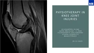
MR APPEARANCE:- Both menisci appear dark , without signal on all sequences. Sag. images ant. & post. horn of MM & LM . Cor. images- meniscal bodies. MM On extreme edge of ant. tibia, trans. lig. joins it superiorly & postly : Arrowhead appearance pointing postly. Lower portion ant. horn. Attachment & upper portion trans. lig. junction.
LM Ant. horn may show striated or speckled, increase SI pattern on Proton density images due to interwining of dense collaginous fibres of ACL with fibrocarti. of LM.
Speckled anterior horn of lateral meniscus
Ant & post horns of LM are equal in size both resemble isosceles triangle. Lig. of Wrisberg seen at its ori. at sup. margin of post horn may appear as round dot adjacent to sup. aspect of post horn & may mimic tear. Due to smaller radius of curvature of LM.: Bow tie configuration is seen.
Cor. cross sections at mid portion of knee : bodies of both menisci seen. Trianguler in shape. Postly cor. cross sections post horns seen as flat bands.
On lat. side, popli tandon courses upwards & lat. at 450. Synovium extends suply & infly about tendon through opening in capsule. Syno. appears increase SI & linear: cor. & sag. images. Med. SI on T1W & spin density images High SI on T2W images.
Transverse lig. Connects ant. horns of both menisci. Thickness : 1 to 4 mm. Arise at ant. sup. portion of ant horn of LM & may appear as small dot ant to meniscus.
ACL Extends from post aspect of med surface of lat.fem. condyle to ant. intercondylar region of tibia. Central support to knee jt. Prevents excessive anterior translation of tibia. Fan shaped tight femoral attachment & wide at tibial attachment 11 mm wide & 38 mm long. ACL is most commonly torn knee lig.
ACL MR APPERARANCE - T2 Sag images. - Straight taut fibres that run parallel to roof of intercondylar notch. - Striated appearance with somewhat high signal near insertion. - Axial images proximally show ACL as a low SI band flattened against med. surface of lat. femoral condyle. Distally, lig. is not well seen. - All imaging sequences demonstrate fat at intercondylar notch, adjacent to ACL origin.
PCL : Lat. surface of med. fem. condyle to post surface of inter condylar region below level of arti. tibial plateau. Lies in sagittal plane & can be seen entirely on a single sag. MR slice. Central support to knee jt. Major stabilizer of the knee. Prevents excessive post translation of tibia,excessive varus & valgus stress & restricts ext. rotation of tibia. 13 mm wide & 38 mm long.
PCL is twice as strong as ACL. Higher tensile strength & so less prone to injury. It is arcuate in shape when knee is extended or slightly flexed & becomes taut with increasing flexion. On MRI sag. images best show PCL: Band like structure of low SI , Arcuate in shape. Nearly horizontal take off at femoral origin & abrupt descent at 450to tibia.
MEDIAL COLLATERAL LIGAMENT :- MCL consist of 3 functional units. 1. Superficial / tibial collateral lig. 2. Deep fibers. 3. Post. oblique fibres. SUPERFICIAL / TIBIAL C L Broad, flat band, 2 cm wide & 2 to 4 mm in thickness. Extend from med. epicondyle of femur to tibia, attaching about 2 cm below jt. line.
DEEP FIBRES=> Function as jt. capsule, attach loosely to periphery of body of med. meniscus. Superior portion constitutes meniscofemoral lig. Firmer attachment to tibia just below jt. line forms coronary lig. POST. OBLIQUE FIBRES=> Blend into post. capsule of knee.
MR APPEARANCE=> TIBIAL COLLATERAL LIG. Best seen on coronal images: homogenously low SI structure on all pulse sequences. Moderately increase SI between superficial & deep fibres.
LATERAL / FIBULAR COLLATERAL LIG. Tubular cord arising from lat. epicondyle of femur to fibular head, where it joins which biceps femoris insertion as conjoined tendon. MR APPEARANCE => Best seen on coronal images: uniformly low SI cord on all pulse sequences.
BONE & BONE MARROW:- Cortical bone of distal femur, proxi tibia & patella : low SI on all pulse sequences. SI of medullary canal reflects changing amounts of haematopoitic & fatty bone marrow with age. Fatty marrow increases with age. Epiphyseal portions of distal F & proxi. T and epiphysoid patella contain fatty marrow. Sensitivity of MRI to bone marrow alterations makes it ideal for detecting traum. injuries.
MUSCLES & TENDONS :- 10 ms & their tendons cross knee jt. Ant.: Quadriceps => Rectus femoris Vastus medialis V. Intermedius V. Lateralis Quadriceps insert on proxi. pole of patella. Anterolat: Tensor fascia lata & its iliotibial band insert at Gerdy s tubercle at anterolat tibia. Post lat.: biceps femoris inserts with fibular CL as conjoined tendon at fibular head. Post med.: sartorius, gracilis & semitendinosus insert at post med. tibia. Semimemdranosus inserts seperately slightly more posteriorly.
Med. & lat. Heads of gastrocnemius arise at post. Supracondy region of F. Popliteal MS belly rests at post. proxi. tibia. Its tendon courses intra-articularly through post horn of LM to popli groove of F. Plantaris ms ori. at postlat. supracondy F & its tendon (longest in body) courses toward ankle.
EXTENSOR MECHANISM=> Quadriceps ms & tendon Patellar lig. Components of patellofemoral jt.
QUADRICEPS TENDON=> Rectus F, V intermedius, V med. & V lat. ms. converge to insert as Q tendon at sup. pole of patella. Fasciae of 4 ms combine to varying degrees to form a multilayered str. Usually V. med. & V. lat. Merge: 3 layered Q tendon with fascia from R. fem. ant. & F from V . int. postly. Also F. of V. med. & lat. combine with each other & with F of either R. fem. or V. int. ms: 2 layered tendon Laminar str. Seen on axial & sag. MRI scans. Partial ruptures of Q tendon involve one of these fascial groups.
PATELLAR LIG. => Extends from distal pole of patella to ant. tibial tubercle. Uniformly low SI best seen on sag. images.
PATELLOFEMORAL JOINT=> Patellar tracking abnormalities imaged with MR P-F joints during incremental knee flexion.
ARTICULAR CARTILAGE :- MRI best suited for evaluation of articular cartilage, different pulse sequences required. Low to intermediate SI on T1W, spin density & T2W images. T1WI intermediate SI compared to low SI of cortex and menisci.
