MR Spectroscopy: Types, Techniques, and Metabolites
Explore the world of MR Spectroscopy, covering single voxel and multivoxel types, important techniques like water suppression and saturation bands, as well as key metabolites and their significance in various conditions. Discover how MR Spectroscopy aids in brain tumor diagnosis and treatment planning.

Download Presentation

Please find below an Image/Link to download the presentation.
The content on the website is provided AS IS for your information and personal use only. It may not be sold, licensed, or shared on other websites without obtaining consent from the author. If you encounter any issues during the download, it is possible that the publisher has removed the file from their server.
You are allowed to download the files provided on this website for personal or commercial use, subject to the condition that they are used lawfully. All files are the property of their respective owners.
The content on the website is provided AS IS for your information and personal use only. It may not be sold, licensed, or shared on other websites without obtaining consent from the author.
E N D
Presentation Transcript
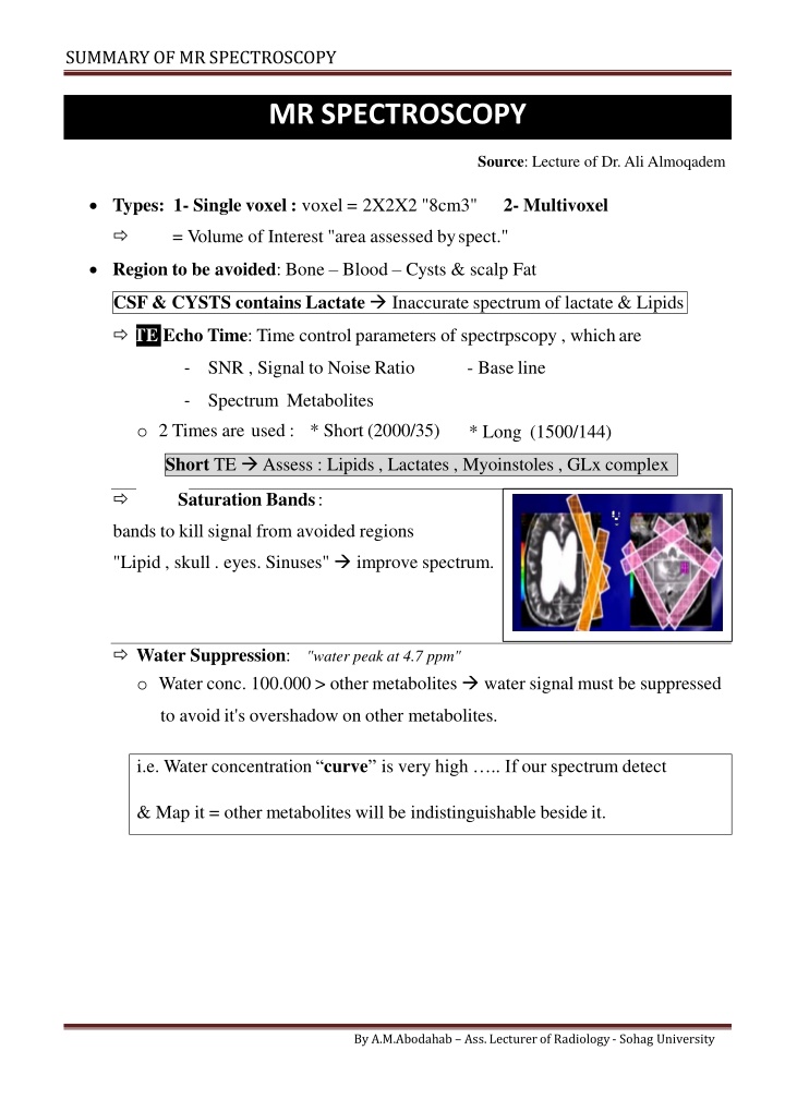
SUMMARY OF MRSPECTROSCOPY MR SPECTROSCOPY Source: Lecture of Dr. Ali Almoqadem Types: 1- Single voxel : voxel = 2X2X2 "8cm3" 2- Multivoxel VOI = Volume of Interest "area assessed byspect." Region to be avoided: Bone Blood Cysts & scalp Fat CSF & CYSTS contains Lactate Inaccurate spectrum of lactate & Lipids TE Echo Time: Time control parameters of spectrpscopy , whichare SNR , Signal to Noise Ratio - Baseline - Spectrum Metabolites - o 2 Times are used : * Short (2000/35) * Long (1500/144) Short TE Assess : Lipids , Lactates , Myoinstoles , GLx complex SATs Saturation Bands: bands to kill signal from avoided regions "Lipid , skull . eyes. Sinuses" improve spectrum. Water Suppression: o Water conc. 100.000 > other metabolites water signal must be suppressed "water peak at 4.7 ppm" to avoid it's overshadow on other metabolites. i.e. Water concentration curve is very high .. If our spectrum detect & Map it = other metabolites will be indistinguishable besideit. By A.M.Abodahab Ass. Lecturer of Radiology - Sohag University
SUMMARY OF MRSPECTROSCOPY METABOLITES & THERE SIGNIFICANCE Some are the main i.e. Stars in every Curve (Cho , Cr , NAA &MI.) Others are seen in certaindiseases. ABC .. When Cho > Cr & NAA = Disease After 2 y of age Spectrum as inAdult AGE VARIATION Neonates Gradual increase Neuronal Maturation High High Adult Diminished NAA Cho. mi Cr. Elevated Increased By A.M.Abodahab Ass. Lecturer of Radiology - Sohag University
SUMMARY OF MRSPECTROSCOPY Metabolites of MR SPECTROSCOPY Phys. Significance Metabolite Locate / ppm Absent in Increased in Diminish in Rare N-Acetyl Aspartate NAA -Marker on Neuron Health -seen only inneuronal tissue -Stable in many Dis. -Used as control -Energy Metabolism Neuronal Damage Tissue of no neurons (Mets/Meningioma) "Canavan's Dis." 2.02 Creatine Cr Trauma Hyperosmolar Hypoxia Tumors 3.03 3.94 Choline Cho - Marker of memb. Synthesis & number of cells Active Tumor Inflammations MS ChronicHypox. NecroticTumor Stroke Abscess In aggressive diseaseprocess 3.2 Lactate Lac Double peak Normally absent or very low -Product of anaerobic metabolism -Released in cell destruct -May due to contamination of scalp Lipids Normally absent 0.9:1.2 3.56 -only short TE -Astrocytes Marker Myoinositol ml Alzheimer Demylenation Low g Glioma GliomatosisC. Tumefactive MS Hepatic Enceph. Sever Hypoxia Hepatic encephalo- pathy 2.1:2.5 -only short TE -Close to each=Glx -Regulate neurotransmitter activities Glutamate & Glutamine Glu Gln OTHERS Succinate = 2.4 Acetate = 1.9 Amino Acids = 0.9 Pyogenic Abcs. Alanine Meningioma By A.M.Abodahab Ass. Lecturer of Radiology - Sohag University
SUMMARY OF MRSPECTROSCOPY SPECTROSCOPY & BRAIN TUMORS - MRS is important aiding tool in diagnosis , but not alone MRS is helpful in differentiating : o Tumor / Tumorlike o Grading cerebralGlioma o TTT planning ofGliomas o Residual / Recurrenttumors o Radiation Injury MRS helping in Grading, . but not Grading Tumoralone Values are slightly different from Paper to Other. By A.M.Abodahab Ass. Lecturer of Radiology - Sohag University
SUMMARY OF MRSPECTROSCOPY PyogenicAbscess Canavan'sDisease EpendymomaRecurrence MassiveInfarction Radiation Injury GlioblastomaMultiform Curve Of Peri LesionVoxel Raised Cho > NAA i.e. ratio > 1 Invasive edge= 1 ry Neoplasm By A.M.Abodahab Ass. Lecturer of Radiology - Sohag University


