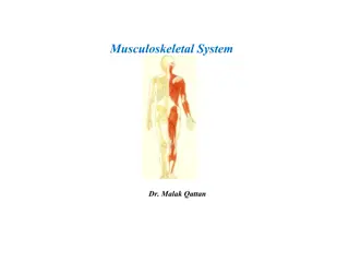
Musculoskeletal System: A Comprehensive Guide
Explore the intricate workings of the musculoskeletal system, encompassing bones, muscles, cartilage, tendons, ligaments, joints, and connective tissues. Discover the vital functions of this system in providing support, stability, movement, and protection to the body. Delve into the human skeleton, bone classifications, and the role of bone marrow in maintaining skeletal health.

Download Presentation

Please find below an Image/Link to download the presentation.
The content on the website is provided AS IS for your information and personal use only. It may not be sold, licensed, or shared on other websites without obtaining consent from the author. If you encounter any issues during the download, it is possible that the publisher has removed the file from their server.
You are allowed to download the files provided on this website for personal or commercial use, subject to the condition that they are used lawfully. All files are the property of their respective owners.
The content on the website is provided AS IS for your information and personal use only. It may not be sold, licensed, or shared on other websites without obtaining consent from the author.
E N D
Presentation Transcript
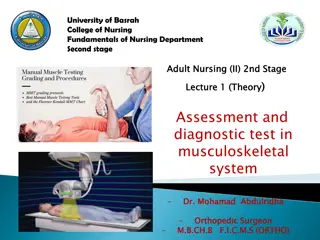
Nursing Care for Patients with Musculoskeletal Problems. 21/10/1446 Dr: Nagwa M., Ahmed
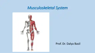
Introduction: A musculoskeletal system (also known as the locomotor system) is an organ system that gives human the ability to move using the muscularand skeletal systems. The musculoskeletal system provides form, support, stability, and movement to the body. It is made up of the: Body's bones (the skeleton). Muscles. Cartilage. Tendons. Ligaments. Joints. Connective tissue, that supports and binds tissues and organs together. 21/10/1446 Dr: Nagwa M., Ahmed
Introduction: The include supporting the body, allowing motion, and protecting vital organs. musculoskeletal system's primary functions The skeletal portion of the system serves as the main storage system for calcium and phosphorus and contains critical components of the hematopoietic system. Bones are connected to other bones and muscle fibers viaconnective tissuesuch as tendonsand ligaments. 21/10/1446 Dr: Nagwa M., Ahmed
Introduction: The bones provide the stability to a body in analogy to iron rods in concreteconstruction. Muscles keep bones in place and also play a role in movement of the bones. To allow motion, different bones areconnected by joints. Cartilage prevents the bone ends from rubbing directly on to each other. Muscles contract (bunch up) to move the bone attached at the joint. 21/10/1446 Dr: Nagwa M., Ahmed
Introduction: Human Skeleton: The Skeletal System serves many important functions: It provides the shapeand form for the body. and theskull and the lungs are protected by the rib cage) Supporting protecting. (brain is protected by Allowing bodily movement. Producing blood for the body. Storing minerals (as calcium and phosphorus) 21/10/1446 Dr: Nagwa M., Ahmed
Human Skeleton: Introduction: 21/10/1446 Dr: Nagwa M., Ahmed
Introduction: Human Skeleton: Humans are born with about 300 to 350 bones; however, many bones fuse together between birth and maturity. As a result an average adult skeleton consists of 206 bones. There are fivegeneral classifications of bones: Long bones. Short bones. Flat bones. Irregular bones. Sesamoid bones. 21/10/1446 Dr: Nagwa M., Ahmed
Introduction: Bone Marrow: Located in long bones Two distinctions of bone marrow (yellow and red). Theyellow marrow: Has fatty connective tissue and is found in the marrow cavity. During starvation, the body uses the fat in yellow marrow forenergy. The red marrow: Is an erythrocytes, platelets, and most leukocytes), approximately 2.6 million red blood cells per second are produced in order to replace existing cells that have been destroyed by the liver. important site for blood cell production (all 21/10/1446 Dr: Nagwa M., Ahmed
Introduction: Muscle: Types of muscle and theirappearance: Cardiac. Skeletal. Smooth. Smooth muscles: Used to control the flow of substances within the lumens of holloworgans. Are notconsciously controlled. 21/10/1446 Dr: Nagwa M., Ahmed
Introduction: Muscle: Skeletal muscles: Have striations that are visible under a microscope due to the components within theircells. Only the skeletal muscles can move the body. Areattached to bones and arranged in opposing groups around joints. Cardiac muscles: Have striations that are visible under a microscope due to the components within theircells. Are found in the heartand are used only to circulate blood Are not under conscious control. Only skeletal and smooth muscles are part of the musculoskeletal system. 21/10/1446 Dr: Nagwa M., Ahmed
Introduction: Tendon: A tough, flexible band of fibrous connective tissue that connects muscles to bones. As muscles contract, tendons transmit the forces to the rigid bones, pulling movement. Tendons can stretch substantially, allowing them to function as springs during locomotion, thereby saving energy. on them and causing 21/10/1446 Dr: Nagwa M., Ahmed
Introduction: Joints: Joints are structures that connect individual bones and may allow bones to move against each other to cause movement. Synovial joints: Joints thatare notdirectly joined. Are lubricated by a solution called synovial Fluid that is produced by the synovial membranes. This fluid lowers the friction between the articular surfaces and is kept within an articular capsule, binding the joint with its taut tissue. 21/10/1446 Dr: Nagwa M., Ahmed
Introduction: Synovial joints: Joints thatare notdirectly joined. Are called produced membranes. lubricated synovial by Fluid the a solution that synovial is by This the friction between the articular surfaces and an articular capsule, binding the joint with its taut tissue fluid lowers is kept within 21/10/1446 Dr: Nagwa M., Ahmed
Introduction: Ligaments: A small band of dense, white, fibrous elastic tissue. Ligaments connect the ends of bones together in order to form a joint. ligaments limit dislocation, movements that maycause breaks. Most or prevent certain Since they are only elastic they increasingly lengthen when under pressure. When this occurs the ligament may be susceptible to break resulting in an unstable joint. 21/10/1446 Dr: Nagwa M., Ahmed
Introduction: Bursa: A small fluid-filled sac made of white fibrous tissue and lined with synovial membrane. Bursa may also be formed by a synovial membrane that extends outside of the joint capsule. It provides a cushion between bones and tendons and/or muscles around a joint; bursa are filled with synovial fluid and are found around almost every major joint of the body. 21/10/1446 Dr: Nagwa M., Ahmed
Assessment: Nursing history: Determine if client is involved in competitivesports and physical condition. History for alcohol use, cigarette smoking, diet, calcium intake < 500 mg daily, age 45, and family history of osteoporosis orotherdiseases. Ask client to describe history of alteration in bone, muscle, or join function. Assess for Hx of pain. Assess normal activity pattern (type of exercise routinely performed). Determine how alteration influences ability to perform activities of daily living (bathing, feeding) and social functions (work, recreation, sexual activities). Assess height loss (firstclinical sign of osteoporosis). 21/10/1446 Dr: Nagwa M., Ahmed
Assessment: Nursing Assessment: Theassessment focuses on: Determining rangeof joint motion. Muscle strength and tone. Joint and muscle condition. Notes in assessment: Assess patient when hewalks, moves in bed, ordo any type of activity. Muscular disorders are manifestations of neurological disease (neurological assessment is performed at the same time). The examination uses inspection and palpation, muscles and joints should be exposed and free to move. Depend on the muscle assessed, the client placed in a sitting, supine, prone, or standing position. 21/10/1446 Dr: Nagwa M., Ahmed
Assessment: Inspection for: Gait (a way of a walking). Posture (anterior, posterior, and lateral): Normal standing posture is an upright stance and parallel alignmentof the hips and shoulders. Common postural abnormalities include: Lordosis (increased lumbarcurvature). Kyphosis (increased thoraciccurvature, olderadult). Scoliosis (lateral spinal curvature). 21/10/1446 Dr: Nagwa M., Ahmed
Assessment: Lordosis (increased lumbar curvature). Kyphosis thoracic curvature, older adult). Scoliosis spinal curvature). (increased (lateral 21/10/1446 Dr: Nagwa M., Ahmed
Assessment: Inspection for: Inspect extremities for size, gross deformity, bony enlargement, alignment, and symmetry (should be bilateral symmetry). 21/10/1446 Dr: Nagwa M., Ahmed
Assessment: Palpation: Apply gentle palpation to all bones, joints, and a rounding muscle in a complete examination noting: Heat. Tenderness. Edema. Resistance to pressure. 21/10/1446 Dr: Nagwa M., Ahmed
Assessment: Range of motion (ROM): All ROM must be performed and noteany limitation in movement. A goniometer instrument used to measure the degree of motion of the particular joint 21/10/1446 Dr: Nagwa M., Ahmed
Assessment: Range of motion (ROM): Normal: no discomfort, firm muscles, no hotness or edema. Full range of motion without anydiscomfort. 21/10/1446 Dr: Nagwa M., Ahmed
Assessment: Muscle tone: Muscle tone: Slight muscular resistance felt by the examiner as relaxed extremity is passively moved through its range of motion. Normal tone is mild. Increase (resistance increased) is called hypertonicity. Hypotonicity: a decrease in muscle tone. muscle tone 21/10/1446 Dr: Nagwa M., Ahmed
Assessment: Muscle Strength: Method of assessmentof musclegroupsstrength: Muscle group Method of assessment Place hand firmly against client's upper jaw. Ask client to turn head laterally against resistance. Place hand over midline of client's shoulder, exerting firm pressure. Have client raise shoulders against resistance. Neck (sternnocleidomastoid) Shoulder (trapezius) 21/10/1446 Dr: Nagwa M., Ahmed
Assessment: Muscle Strength: Method of assessmentof musclegroupsstrength: Muscle group Elbow : a. BICEPS: b. TRICEPS: Method of assessment a. Pull down on forearm asclient attempts to flex arm. b.As client's arm is flexed, apply pressure against forearm.Ask client to straighten arm. A.When client is sitting, apply downward pressure to the thigh.Ask client to raise leg up from the table. B. Client sits, holding shin of flexed leg. Ask client to straighten leg against resistance Hip: A. quadriceps: B. gastrocnemius: 21/10/1446 Dr: Nagwa M., Ahmed
Assessment: Muscle Strength: Muscle strength. Grading scale. Muscle function level Grade No evidence of contractility 0 Slight contractility, no movement 1 Full range of motion, passive movement 2 Full range of motion with gravity 3 Full range of motion against gravity (some resistance) 4 Full range of motion against gravity (full resistance) 5 21/10/1446 Dr: Nagwa M., Ahmed
Laboratory Tests: Antinuclearantibodies: Result either positive or negative (normal) and if positive indicate autoimmune disease. C-reactiveprotein: Protein appears in inflammatory cases, require fasting and noteany medication used. Normal Result is negative. 21/10/1446 Dr: Nagwa M., Ahmed
Laboratory Tests: Erythrocytesedimentation rate (ESR): in inflammatory, conditions. Increased infections, and cancerous Performed within 3 hours after blood drawing and you should noteany medication and pregnancyor menstruation. Normal in male up to 20 mm/h and in female up to 30 mm/h. 21/10/1446 Dr: Nagwa M., Ahmed
Laboratory Tests: Rheumatoid factor: IgM antibody developed against IgG. Normally test is negative. Uricacid: Elevated in gout. Serum: male 2.1-8.5 mg/dl. Female: 2-6.6 mg/dl Urine: 250-750mg/24h. 21/10/1446 Dr: Nagwa M., Ahmed
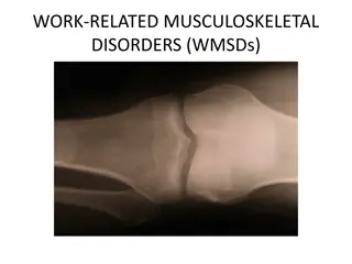
Radiologic Studies: Computed tomography (CT scan). Magnetic resonance imaging (MRI). X-ray. Arthrography/Arthrogram: Test includes injection of radiopaque dye into the joint then visualization of the joint taken place. 21/10/1446 Dr: Nagwa M., Ahmed
Radiologic Studies: Arthrography/Arthrogram: Nursing responsibility: Gathering the equipment and needed materials (sterile equipmentsand local anesthesia) Explain theprocedure to theclient. Obtain consent form. Check thesite foredemaand increased pain. Administeranalgesia. Apply elastic bandage. 21/10/1446 Dr: Nagwa M., Ahmed
Electromyography: Measuring the muscle activity at rest and during voluntary muscle contraction byadministering a needle electrode into the muscle. Used to detect primary musculardisorders. Testwill take 1 h. Nursing responsibility: Explain the procedure to the client. Obtain consent form. Prohibit smoking and caffeineat least 3 h before test. Observe the site for hematomaand inflammation. 21/10/1446 Dr: Nagwa M., Ahmed
Arthroscopy: Directvisualization of the joint. Done in operation room under local orgeneral anesthesia. Nursing responsibility: Aftertest frequently check the neurovascularof theexamined part. Elevate theclient leg. Apply compression dressing. Administeranalgesia. 21/10/1446 Dr: Nagwa M., Ahmed
Arthrocentesis: Sterile aspiration of the fluid found in the joint after local anesthesia. Used inflammation. to administer medication and detect infection or Nursing responsibility: Explain the procedure to theclient. Obtain consent form. Apply pressureand ice. Check the site foredemaand increased pain. 21/10/1446 Dr: Nagwa M., Ahmed
Musculoskeletal Changes Associated with Age: The following changes occur with age: Decreased muscle mass and strength. Bone demineralization (more pronounced in women). Shortening of trunk as result of intervertebral space narrowing. Decreased joint mobility and rangeof joint motion. 21/10/1446 Dr: Nagwa M., Ahmed
Musculoskeletal Changes Associated with Age: Nursing care: Help in self-care procedure and otheractivity. Prevent fractures by ensuring safe hospitalization, home, and community places. Teach client use of thewalking aides. Provide psychological support. Educate client range of motion and other suitable exercises with adequate period of rest. Educate client about the suitablediet. 21/10/1446 Dr: Nagwa M., Ahmed
Surgical Intervention for Problems of Musculo- skeletal System: Joint Replacement(Arthroplasty): Is total or partial replacement of the joint with a synthetic prosthesis. Used mainly in severe chronic arthritis and extensive joint trauma to: Restores mobility and stability. Relieves pain. Increased senseof independenceand self-worth. 21/10/1446 Dr: Nagwa M., Ahmed
Surgical Intervention for Problems of Musculo- skeletal System: Joint Replacement(Arthroplasty): Complications: Infection (removed). Hypovolemic shock. Fat embolism. Thromboembolism. Pulmonary embolism (most common cause of death) and pneumonia. Compression of the nervesand vascular necrosis. Dislocation or loosening of the prosthesis. 21/10/1446 Dr: Nagwa M., Ahmed
Surgical Intervention for Problems of Musculo- skeletal System: Joint Replacement(Arthroplasty): Nursing interventions: Before surgery: Preparation which include testing, examination, and teaching (mainly by doctor). Prepare the patient for an extended period of rehabilitation (physical therapist). Inform patient that he experience pain and may worsen for several weeks (use of analgesics). Ensure that the patient or family member has signed a consent form. Ensure that other requirements of operation are ready such as prosthesis and blood if needed. Provide psychological support. 21/10/1446 Dr: Nagwa M., Ahmed
Surgical Intervention for Problems of Musculo- skeletal System: Joint Replacement(Arthroplasty): Nursing interventions: After surgery: Keep clienton bed rest for the prescribed period. Maintain theaffected joint in properalignment. Assess the patient's level of pain and provide analgesics as ordered. Monitor for complications of joint replacement (assess vital signs, neurovascular and motor status distal to the site of joint replacement and skin frequently). Watch for signs of a fat embolism (fever, disorientation, dyspnea, tachypnea, sleepiness, agitation, coma, and death mayoccur). 21/10/1446 Dr: Nagwa M., Ahmed
Surgical Intervention for Problems of Musculo- skeletal System: Joint Replacement(Arthroplasty): Nursing interventions: After surgery: Change position the patient (to enhance comfort and prevent pressuresores). Encourage coughing and deep breathing and adequate fluid intake. Administerprophylacticantibiotics. Patient will be out of bed the 1st or 2nd day after surgery (follow thesurgeon instructions). Encourageclient to follow the physical therapists instructions. Encourageclient to take the prophylacticantibiotic in home. 21/10/1446 Dr: Nagwa M., Ahmed
Surgical Intervention for Problems of Musculo- skeletal System: Amputation: Amputation is the removal of the limbor part from it. Used mainly in gangrene, cancer, trauma, and vascular diseases. Purpose: Preserve function in a remaining part. Preventdeath. 21/10/1446 Dr: Nagwa M., Ahmed
Surgical Intervention for Problems of Musculo- skeletal System: Amputation: Complications: Infection. Contractures in theremaining limb part. Skin breakdown. Phantom pain is a sensation of pain (itching, or numbness in thearea of amputation) developsafter 2 to 3 months. Depression. 21/10/1446 Dr: Nagwa M., Ahmed
Surgical Intervention for Problems of Musculo- skeletal System: Amputation: Nursing interventions: Before surgery: Answerany questions the patient may have. Prepare the patient for surgery and providing care and instruction thatwill be taken after theamputation. Provide emotional support (loss of dependence). Arrange forsupportgroup (with same problem) meeting. Explain that the patient may "feel" phantom pain. As ordered, administerantibioticsand premeditations. Consent form and otherpreparations. a body part and 21/10/1446 Dr: Nagwa M., Ahmed
Surgical Intervention for Problems of Musculo- skeletal System: Amputation: Nursing interventions: After surgery: Monitorvital signs. Check dressings frequently and change them as ordered. Assess for bleeding (through thedressing). Elevate the limbon a pillow orothersupport (If ordered). Assess drain patency (working) and note the amount and characterof drainage. 21/10/1446 Dr: Nagwa M., Ahmed
Surgical Intervention for Problems of Musculo- skeletal System: Amputation: Nursing interventions: After surgery: Keep drainage bag below the level of the body and empty it after it become filled with fluid and after informing physician. Noteamount, color, consistency. Inform physician if drainage system drain large fresh blood and if oozing. Assess for pain and provide analgesics (give analgesics about 30 minutes beforescheduled exercises orambulation). Care forcast (if applied). Maintain proper body alignment and regular physical therapy (passive ROM exercises). 21/10/1446 Dr: Nagwa M., Ahmed
Surgical Intervention for Problems of Musculo- skeletal System: Amputation: Nursing interventions: After surgery: Teach activities to "strengthen" the residual limb. give the patient informationaboutavailable prostheses Encourage the patient to adopt a positive outlook (spiritual counselor). Instruct the patient to examine the limb daily for swelling, redness, orexcessive drainage, skin changes, and pain. Instruct the patient about good hygiene (wash the limb daily at night with mild soap and water and then to rinse and to gently dry it) and bandage itwhen dry. 21/10/1446 Dr: Nagwa M., Ahmed
Traumatic Injuries: Contusion (bruise): Contusion is the most common and is the result of an injury to the soft tissues. The injury causes a rupture of small blood vessels and subsequent bleeding into (discoloration: black-and-blue) or hematoma. tissues ecchymosis Treatment: Elevation of the affected part. Application of cold immediately and if no bleeding after 24 hours, heat may be applied to relieve muscle soreness. 21/10/1446 Dr: Nagwa M., Ahmed
Traumatic Injuries: Sprains: Sprain involves ligaments, tendons, and muscles and occurs as the result of twisting of a joint beyond its normal range of motion Ligaments are torn. Tendons may be pulled from the bone. Blood vessels are ruptured, which causes contusions with ecchymosis Pain related topressureon nerveendings. Disturbance of circulation and lymph drainage muscle spasm & edema. 21/10/1446 Dr: Nagwa M., Ahmed