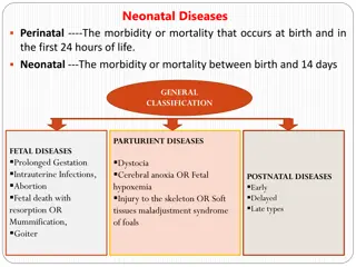
Neonatal Hypoglycemia and Its Management
Neonatal hypoglycemia is a common metabolic issue in newborns, often associated with endocrine disorders and possible neurological effects. Glucose plays a vital role in the energy supply to fetuses and newborns, with certain factors predisposing infants to hypoglycemia. Early detection and intervention are crucial in preventing long-term complications. Learn about the clinical definitions, intervention thresholds, and management strategies for neonatal hypoglycemia in this comprehensive guide.

Download Presentation

Please find below an Image/Link to download the presentation.
The content on the website is provided AS IS for your information and personal use only. It may not be sold, licensed, or shared on other websites without obtaining consent from the author. If you encounter any issues during the download, it is possible that the publisher has removed the file from their server.
You are allowed to download the files provided on this website for personal or commercial use, subject to the condition that they are used lawfully. All files are the property of their respective owners.
The content on the website is provided AS IS for your information and personal use only. It may not be sold, licensed, or shared on other websites without obtaining consent from the author.
E N D
Presentation Transcript
Dr v sowjanya Bhanu Senior resident
Hypoglycemia is one of most commonmetabolic problem in neonates. Persistent hypoglycemia is most likelyassociated with endocrine disorders and possible neurologic sequelae
Glucose provides 60-70% of energy to fetus and newborn. Fetal glucose is approximately 2/3 ofmaternal levels.(Trans placental facilitated diffusion) Why prone to develophypoglycemia:- 1.Umblical cord cutting at birth. 2. Inadequate storage of glycogen. 3. Immature adaptive mechanisms. 4. Prone to long term neurological damage.
Lowest value upto 25-40 mg/dl in the first1 -2 hours of life. Stabilize by 65-70 mg/dl by 3-4 hours of age spontaneously or with feed/intervention
Incidence: 1.3-3/1000 livebirth. Incidence varies with definition, method, timing of feed and type of glucose estimation method. Plasma value is 10-15% higher than bloodglucose value. Incidence is higher in at riskneonates (LPT>SGA>IDM>LGA) population,
Clinical definition:- Based on symptoms associated with hypoglycemia and resolution of symptoms when glucose restored in normal range. Sometimes development of signs and symptoms may be late.
Glucose level at which intervention should be considered. It is an indication of action not diagnostic of disease or abnormality. It is based on clinical experience and analysis of available evidences.
Healthy term: <24 hrs: 30-35 mg/dl at one time 45mg/dl if persist after feeding or recurs. >24 hrs: 45-50 mg/dl Symptomatic: 45 mg/dl Asymptomatic with risk factor: 36 mg/dl Any baby with <20-25 mg/dl
LBW infants(<2.5 kg) Preterm (<35weeks) SGA (BW<10thcentile),L G A, I D M Rh hemolytic disease Post exchange transfusion Infants on TPN Sick neonates(sepsis, shock, asphyxia, polycythemia, distress etc.) Mother receiving terbutaline, labetalol, oral hypoglycemics
Category of infants At risk neonates 2. Sick infants 3. Stable VLBW4. 4. infants on TPN 5. IDM Time schedule At 2, 6, 12, 24, 48, 72hrs Every 6-8 hrly Initial 72 hr : 6-8hrly After 72 hrs: Once a day At 1, 2, 3, 6, 12, 24,26 and 48 hrs 1 hr after exchange 1. 6. Exchange transfusion
Hyperinsulinemic hypoglycemia: Major cause of persistent and recurrent hypoglycemia IDM: historically known mc cause Congenital/genetic: Mutation of pancreatic beta cell ATP sensitive K+ channel . (ABCC8,KCNJ11 Encodes for SUR1,Kir )
Birth asphyxia, Development syndromes- Beckwith-wiedemann syndrome Erythroblastosis Maternal tocolytic drugs Malpositioned umbilical artery catheter Abrupt cessation of high glucose infusion. After exchange transfusion with high BG concentration. Insulin producing tumors eg. nesidioblastosis, islet cell adenoma or dysmaturity.
Prematurity IUGR Inadequate caloric intake Delayed onset of feeding
Peri-natal stress Exchange transfusion (reactive hypoglycemia Endocrine deficiency Defect in carbohydrate and AA metabolism Polycythemia Maternal therapy with beta blocker causes prevention of sympathetic stimulation of glycogenolysis
SYMPTOMS: Irritability Tremors Jitteriness Exaggerated Moros reflex Seizures,high pitched cry Lethargy,hypotonia,cyanosis Poor feeding
Reagent strips/Glucometer:- Most widely used method, mainly used for screening Measures whole blood glucose level which is 15% lower than plasma value. Unreliable at the lower values. Capillary samples ( Heel prick).
Glucose oxidase (colorimetric)method:- Used in laboratories Most reliable and accurate method Glucose electrode ( Blood gasanalyser):- Minimal volume of blood required
Goal of current management is to anticipate a n d prevent hypoglycemia rather than treatment. Asymptomatic:-BGL 20-45mg/dl: Trial of feed and repeat BGL after 1 hr If repeat BGL >45, 2 hrly feed with 6 hrly BGL for 48 hrs. If repeat BGL <45, confirm with lab report and management is as for symptomatic hypoglycemia
BGL <20mg/dl: Start treatment with IV glucose Goal: >45 in first 24 hr and >50 thereafter.
Symptomatic 1. Urgent treatment: 2 ml/kg of 10% D/W (200mg/kg) over 1 minute 2. Continuing therapy: Start GIR @6-8 mg/kg/min
Recheck BGL 15-30 minute after bolus, then hrly until stable Additional bolus of 2 ml/kg may be needed If BGL is stable & in normal range, feeding may be started & GIR may be tapered.
Refractory: GIR requirement of >12mg/kg/min for >24 hrs. Prolonged/Persistent: Unstable BGL beyond5 7 days. Causes:- Hyperinsulinemic hypoglycemia Congenital hypopituitarism, hypothalamic deficiency Adrenal insufficiency, epinephrine deficiency GSD, galactosemia, fructose intolenrance Defects in AA metabolism eg-MSUD, tyrosinemia Polycythemia
Critical lab sample: Evaluation requires drawing blood for insulin ,cortisol,aminoacids when glucose levelis <40mg/dl Glucose Insulin (>2 micro U/ml diagnostic) I:G ratio during hypoglycemia >0.3-0.5 Beta hydroxy butyrate and FFA levels Cortisol: to screen the integrity of HPA axis
If insulin level is normal for blood glucose consider additional testing: GH, ACTH, T4 &TSH, Glucagon, Cortisol Plasma AA, Blood NH3, Blood lactate level Urine ketones, reducing substance, AA, organic acids Genetic testing for mutations 18F fluoro L-DOPA PET scan to identify focal lesion in pancreas to consider for subtotal pancreatectomy.
Hydrocortisone: 5 mg/kg/day iv q 12hrly Peripheral glucose utilization, gluconeogenesis and effect of glucagon. Glucagon: 0.025-0.2 mg/kg iv/im/sc. Max- 1mg. Increase glucose release Temporary measure and can be used in infants with good glycogen stores. Diazoxide: 5-8 mg/kg/day q 8-12hrly. Inhibit insulin release Octreotide: 5-20 mcg/kg/day iv/sc q 6-8hrly. Inhibit insulin secretion, can be used when diazoxide does not successfully control BGL. Subtotal pancreatectomy
1. MRI Scan : Typical pattern of CNS injury particularly in parieto-occipital cortex and sub cortical white matter. 2. Neonates have developmental delay, cerebral palsy, motor impairment, blindness and hearing impairement.. 3. Babies who have had symptomatic hypoglycemia should have close follow up at 3, 6, 9, 12 & 18 month age for growth, neurodevelopment, vision and hearing loss.
DEFINITION: Hypocalcemia is defined as total serum calcium of less than 7 mg/dL (1.75 mmol/L)or ionized calcium less than 4 mg/dL (1mmol/L) In very low birth weight infants the ionised calcium levels of 0.8 to 1mmol/lit are common and not usually associated with clinical symptoms
The early onset hypocalcemia: presents within 72 h requires treatment with calcium supplementation for at least 72 h. late onset hypocalcemia: usually presents after 7 days and requires longer term therapy.
Prematurity Preeclampsia Infant of Diabetic mother Perinatal stress/ asphyxia Maternal intake of anticonvulsants (phenobarbitone, phenytoin sodium) Maternal hyperparathyroidism Iatrogenic (alkalosis, use of bloodproducts, diuretics, phototherapy, lipid infusions etc
Asymptomatic Symptomatic: a) Neuromuscular irritability,Myoclonic jerks Jitteriness,Exaggerated startle Seizures b)Cardiac involvement:Tachycardia,prolonged QT Interval,decreased contractility, c)Other symptoms like apnea,cyanosis,tachypnea,laryngospasm are noted
Laboratory: By measuring total or ionised serum calcium Ionised calcium is preferred mode for measuring hypocalcemia ECG: QoTc>0.22 seconds or QTc>0.45 sec QoT is measured from origin of q to origin of t wave QT is measured from origin of q wave to end of t wave
Bradycardias Arrythmias Skin and subcutaneous tissue necrosis may occur due to extravasation Hepatic necrosis may occur if tip of uvc lies in branch of portal vein
presents at the end of the first wk of life. It is usually symptomatic in the form of neonatal tetany or seizures. This is usually caused by high phosphate intake (iatrogenic).
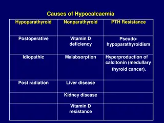
Increased phosphate load Cow milk, renal insufficiency Hypomagnesemia Vitamin D deficiency Maternal vitamin D deficiency Malabsorption Renal insufficiency Hepatobiliary disease PTH resistence Transient neonatal pseudohypoparathyroidism
Hypoparathyroidism Primary Hypoplasia, aplasia of parathyroid glands - (Di George s syndrome), CATCH 22 syndrome ,activating mutations of calcium sensing receptor Secondary Maternal hyperparathyroidism Metabolic Syndromes Kenny-caffey syndrome Long-chain fatty acyl CoAdehydrogenase deficiency Kearns-sayre syndrome
Iatrogenic Citrated blood products, Lipid infusions, Bicarbonate therapy Diueretics, Glucocorticosteriods Phosphate therapy , Alkalosis Phototherapy


")


method:-")